


In the first article of this two-part series discussing tarsal fractures (Perry, 2020), the surgically-relevant anatomy of the tarsus was reviewed, the diagnostic imaging techniques commonly used were evaluated and the diagnosis and treatment of talar fractures was discussed. This article concentrates on the diagnosis and treatment of calcaneal fractures; central tarsal bone fractures; isolated fractures of the numbered tarsal bones; and malleolar fractures.
Fractures of the calcaneus
The calcaneus is the largest and longest bone of the tarsus (Evans, 1993). The distal half of the bone is wide transversely and possesses three facets and two processes whereby it is fitted with the talus to form a very stable joint. The tuber calcanei serves for the insertion of the calcanean tendon. Its slightly bulbous free end contains the medial and lateral processes which are separated by a wide groove. A jutting shelf, the sustentaculum tali, leaves the medial side of the bone. On the plantar side of this process is a wide shallow groove over which the tendon of the flexor hallucis longus glides. On the dorsomedial side is a concave oval facet, the facies articularis talus media, for articulation with the middle articular surface of the talus. The dorsal articular surface, facies articularis talaris dorsalis, is convex, as it articulates with the comparable concave surface of the talus. The most distal and smallest articular surface on the dorsal part of the bone is the facies articularis talaris distalis. This surface is confluent with a small articular facet for the central tarsal on the distal surface. Between the middle and distal articular surfaces is the calcanean sulcus. This sulcus concurs with a similar one on the talus to form the tarsal sinus. On the distal end of the calcaneus is a large flat facies articularis cuboidea, for articulation mainly with the central tarsal bone and by a small facet with the talus (Evans, 1993).
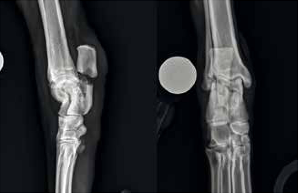
Fractures of the calcaneus that have been described include avulsions of the superficial digital flexor tendon, Salter Harris type 1 or 2 fractures involving the proximal calcaneal physis, mid-body fractures (Figure 1), lateral sagittal slab fractures, dorsomedial slab fractures, fractures of the sustentaculum tali and avulsion fractures of the base of the calcaneus (Dee, 1984a; 1984b; Ost et al, 1987; Denny and Butterworth, 2000). Multiple combinations of fractures have also been seen, with the most frequently reported combination being a fracture of the calcaneal body, a small craniomedial sagittal slab fracture and a lateral sagittal slab fracture (Ost et al, 1987). The fracture fragments in calcaneal fractures are usually significantly displaced, due to the distractive forces applied to the attached tendons and ligaments (Matthiesen, 1983).
Calcaneal fractures are commonly encountered in racing Greyhounds. In one study (Ost et al, 1987), anatomic dissection of two Greyhound tarsi with fractures of the calcaneus revealed that every calcaneal fragment represented an area of origin or insertion of a tendon or ligament. The talocalcaneal ligament inserts at the area where dorsomedial slab fragments occur, the lateral plantar ligament originates from the lateral aspect of the body of the calcaneus (the area of the lateral sagittal fractures). The middle plantar ligament originates from the plantar base of the calcaneus (where the plantarodistal chip fractures or transverse base fractures occur). The medial plantar ligament originates from the sustentaculum tali. The common calcanean tendon attaches to the calcaneal tuber (Ost et al, 1987; Evans, 1993).
Calcaneal fractures can occur with or without other tarsal bone fractures, with the most common concomitant fractures being of the central tarsal bone (Ost et al, 1987). Different pathogeneses have been reported depending on whether the central tarsal bone remains intact or not. When the central tarsal bone is fractured, the talus travels distally and acts as a fulcrum over which the calcaneus fractures. In this situation most calcaneal fractures are either midbody shaft fractures or tarsal ligament avulsion fractures e.g. dorsomedial slabs or lateral sagittal slabs (Ost et al, 1987; Dee, 2005). Calcaneal fractures not associated with central tarsal bone fractures are believed to result from extreme tension on the plantar aspect of the tarsus. The tension is created by the pull of the common calcanean tendon and is transmitted through the calcaneus via the plantar ligaments to the metatarsals. If the tensile forces exceed the strength of the calcaneus, avulsion of the middle plantar ligament occurs, resulting in a transverse fracture or a plantarodistal chip fracture of the base of the calcaneus. Due to loss of plantar support, plantar proximal intertarsal subluxation develops (Ost et al, 1987). Other causes of calcaneal fractures in dogs include pathologic fracture secondary to primary bone neoplasia (Gillick and Galbo, 2003) and fractures secondary to implant placement (Allen et al, 1993; Fettig et al, 2002).
Some differing pathogeneses have been reported in feline patients. Cats are suspected to develop stress fractures of the calcaneus (Cantatore and Clements, 2015) and the calcaneus has also been reported to be one of the bones at risk of fracture in cats with knees and teeth syndrome (KaTS) (Longley et al, 2016; Reyes et al, 2019), so it may be prudent to screen cats with calcaneal fracture for fractures of other bones and dental anomalies upon presentation.
Mid-body calcaneal fractures are common in racing Greyhounds, usually affecting the right side. As stated above, these fractures tend to be associated with fracture of the central tarsal bone or subluxation of the proximal intertarsal joint if the central tarsal bone remains intact (Ost et al, 1987). Options for stabilisation include pins and tension band wires or a laterally applied plate (Denny and Butterworth, 2000). The use of pins and tension band wires has been reported more commonly than plate fixation in the veterinary literature. This is likely because the majority of the literature evaluating calcaneal fractures concentrates on racing Greyhounds and in this population comminuted fractures are less common. However, a recent paper evaluated calcaneal fractures in non-racing dogs (Perry et al, 2017) and reported that comminuted fractures were more common in this population. This is likely due to the fact that calcaneal fractures in non-racing dogs are more likely to occur secondary to traumatic events rather than secondary to a central tarsal bone fatigue fracture. In the study evaluating non-racing dogs, stabilisation with plates and screws appeared to be associated with a better prognosis than treatment using pins and tension bands, screws or combinations of these techniques (Perry et al, 2017). Therefore, plate stabilisation may be worthy of consideration in non-racing dogs with calcaneal fractures (Figures 2 and 3).


For treatment using pins and tension band wires, the patient is placed in sternal recumbency with the leg extended caudally with the tarsus in extension such that the common calcaneal mechanism is relaxed. A lateral plantar approach is followed by a midsubstance incision of the lateral retinaculum of the superficial digital flexor tendon where it attaches to the lateral process of the tuber calcanei. The tendon is then reflected medially, facilitating exposure and reduction of the fracture. Fracture reduction and maintenance is accomplished with the aid of straight Vulsellum forceps. The two sets of paired points are placed at the tuber and at the base of the calcaneus. Traditional management consists of small, paired pins placed as far medially and laterally as possible to minimise the inevitable irritation to the superficial digital flexor tendons. The figure-of-eight wire is placed cranially to the well-seated bent-over pins. The distal aspect of the wire is located in a transosseous tunnel in the base of the calcaneus. Alternatively, single or paired pins respectively may be placed in a mediolateral or craniocaudal plane and countersunk in the tuber to minimise tendon irritation (Dee, 2005). As the calcaneus is a dense bone, it may be necessary to predrill pin holes with a drill bit one size smaller than the anticipated pin size. The superficial digital flexor tendon is reattached before routine closure. Laterally-applied plates and screws can also be placed through a similar approach and for complex cases, supplementing the lateral plate with a tension band wire can be considered (Dee, 2005). Fractures with loss of plantar support require a tension band wire or a neutralisation plate to counteract distracting forces on the plantar surface of the calcaneus (Ost et al, 1987). A modified Robert-Jones dressing is generally applied postoperatively, to minimise soft-tissue swelling (Dee, 2005).
Slab fractures of the distolateral or dorsomedial calcaneus are often complicated by other injuries, for example luxation of the adjacent talus or fracture of the central tarsal bone. Whether seen as an isolated injury, or in conjunction with other injuries, lag screw fixation is used for repair in most of these cases (Denny and Butterworth, 2000; Dee, 2005). For oblique sagittal fractures involving the lateral surface of the long axis of the calcaneus it is generally possible to place at least two lateral-to-medial lag screws (Dee, 2005). If there is comminution then it may be necessary to combine the lag screws with a pin and tension band wire or plate fixation (Denny and Butterworth, 2000).
Salter Harris type 1 or 2 fractures are seen in skeletally immature animals (Dee, 2005). In these cases, the epiphysis is distracted by the gastrocnemius tendon. Kirshner wires and a figure-of-eight tension band wire are generally used for stabilisation (Denny and Butterworth, 2000).
Avulsion fractures of the base of the calcaneus result in plantar instability and subluxation of the proximal intertarsal joint and treatment is generally by arthrodesis of the calcaneoquartal joint (Ost et al, 1987; Denny and Butterworth, 2000).
Stress fractures of the calcaneus in cats may present with either acute, complete calcaneal fracture or alternatively with remodelling or incomplete fractures that progress to complete fractures with time. The bilateral nature and simple similar transverse fracture configurations at the base of the calcaneus, in addition to the absence of trauma in these cases, prompts the suspicion of stress fracture (Cantatore and Clements, 2015). Both of the cats that have been reported with this injury were treated via partial tarsal arthrodesis with lateral plate fixation.
The prognosis following calcaneal fracture in racing dogs is generally good. In one study, 95% of racing Greyhounds treated surgically were sound at the time of radiographic union and 44% returned to racing (Ost et al, 1987). A caveat to this is that in the racing Greyhound, if the repair involves arthrodesis of the proximal intertarsal joint then the animal is unlikely to return to racing form (Denny and Butterworth, 2000). These positive prognoses, however, are all based on literature exclusively evaluating racing Greyhounds. A recent study evaluated calcaneal fractures in non-racing dogs and cats and looked at complications, outcome and associated risk factors (Perry et al, 2017). This study reported a high occurrence of complications associated with calcaneal fracture stabilisation in non-racing dogs and cats, and poorer outcomes were encountered in animals that suffered complications. Based on this study, the positive prognosis associated with calcaneal fractures in racing dogs should be modified when speaking to owners of non-racing dogs with similar injuries.
Fractures of the central tarsal bone
The central tarsal bone lies on the medial aspect of the tarsus, articulating proximally with the talus and calcaneus, laterally with the proximal half of the fourth tarsal bone and distally with the third, second and first tarsal bones. A strong medial plantar ligament runs from the sustentaculum tali, attaching to the plantar process of the central tarsal bone and inserting distally onto the tarsometatarsal joint capsule. The dorsal and intertarsal ligaments are comparatively weak (Evans, 1993).
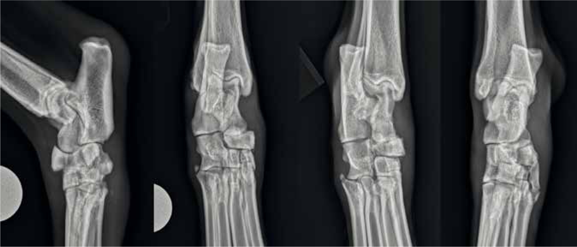
Fracture of the central tarsal bone is the most common tarsal injury in racing Greyhounds and is often associated with concomitant fractures of the calcaneus, fourth tarsal bone, second tarsal bone and the base of the fifth metatarsal bone (Boudrieau et al, 1984a). Fractures of the central tarsal bone have been classified into five types (Dee et al, 1976): type I is a dorsal slab fracture with no displacement; type II is a dorsal slab fracture with displacement (Figure 4); type III is a sagittal fracture with displacement of the medial fragment; type IV have both dorsal and medial slab fractures; and type V is a severely comminuted type IV (Dee, 2005). Type III and IV fractures are the most common in racing Greyhounds, with type IV fractures having an incidence of 68% (Boudrieau et al, 1984a), while type V fractures are most common in non-racing dogs (Armstrong et al, 2019). Boiled-out specimens of type IV fractures show, in addition to the two main bone fragments, multiple small fragments from the body of the bone (Vaughan, 1987). Other injuries reported to involve the central tarsal bone in dogs include dorsomedial luxation leading to partial collapse of the medial aspect of the tarsus (Lorinson and Grosslinger, 2001); fractures of the plantar process of the central tarsal bone together with dorsomedial displacement of the body of the central tarsal bone (Guilliard, 2007); and fracture of the plantar process without luxation of the central tarsal bone (Galateanu et al, 2011). Although rare, central tarsal bone fracture has also been reported to occur in the cat following trauma (Cinti et al, 2016).
The central tarsal bone acts as a buttress supporting the medial side of the tarsus under compression. This compressive force is thought to be greater in the right tarsus when running around an anticlockwise bend (Hickman, 1975; Dee et al, 1976; Dee, 1981). A collapse of the dorsomedial buttress of the bone is a common feature of fracture types III, IV and V with the medial fragment rotating in a medioplantar direction, resulting in tarsus varus and plantar convexity (Guilliard, 2000). Central tarsal bone fractures in racing Greyhounds are considered to be fatigue fractures. Asymmetric cyclic loading induces a significant adaptive micromodelling response in the trabecular bone of the right central tarsal bone of racing Greyhounds as well as subtle changes in the external contours of the dorsal cortex of the bone (Johnson et al, 2000). This response is site-specific, occurring in a region of the right central tarsal bone which is frequently affected by fatigue fracture (Boudrieau et al, 1984a). Fractures occur either at, or adjacent to this sclerotic, dorsal bone, indicating that the mechanism for central tarsal bone fracture in the racing Greyhound may be similar to that of third carpal bone fractures in racehorses (Johnson et al, 2000); fatigue fracture is induced if the magnitude and duration of ongoing cyclic stresses placed upon sclerotic subchondral bone is sufficiently great, with modulus-mismatch leading to concentration of shear stresses and final failure at the interface between dissimilar moduli (Young et al, 1991; Firth et al, 1999).
The specific treatment recommended for the most commonly encountered central tarsal bone fractures will be reviewed individually below, but, in all types of fractures of the central tarsal bone, internal fixation gives superior results to treatment with external coaptation (Boudrieau et al, 1984b). Articular fractures require precise reduction of the articular component and rigid fixation to minimise development of degenerative joint disease and to achieve normal long-term function (Roy and Dee, 1994). External immobilisation of displaced fractures of the central tarsal bone results in healing by intertarsal ankylosis. There may be a deformity of the hock after this treatment and it is certainly not considered ideal; however, many dogs return to functional and comfortable limb use after a period of 3 months (Bateman, 1958).
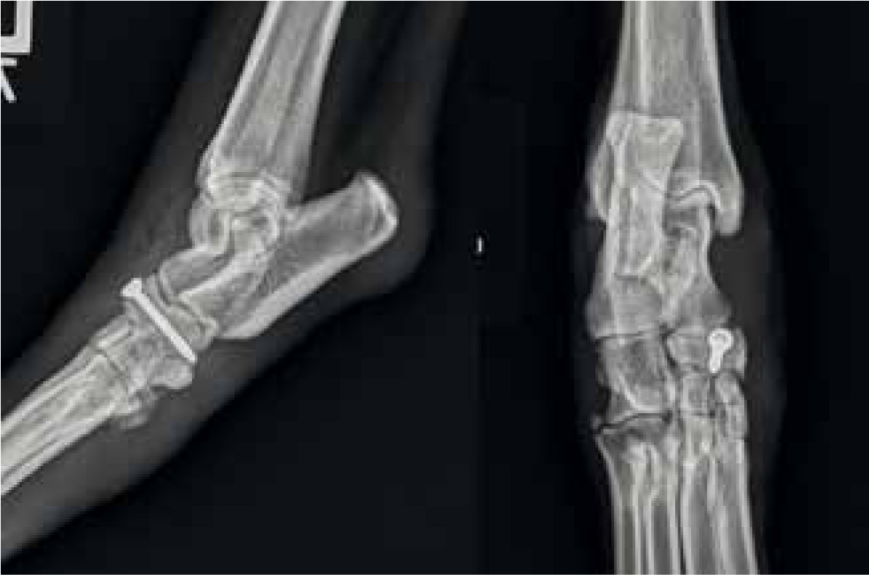
Type II fractures are repaired by the dorsoplantar placement of a lag screw through the two fragments of the central tarsal bone (Figure 5).
Type III fractures are repaired by the mediolateral placement of a lag screw into the fourth tarsal bone (Dee et al, 1976), although an additional screw through the dorsal aspect of the central tarsal bone has also been recommended in case there is a non-displaced dorsal slab fracture that has not been noted radiographically (Boudrieau et al, 1984b).
Type IV fractures are repaired by the use of two lag screws, roughly perpendicular to each other, placed through the medial and dorsal fragments (Dee et al, 1976; Boudrieau et al, 1984b). Many of the smaller fragments found in type IV central tarsal bone fractures are articular, and precise reduction is only attempted on the two large components; the smaller fragments are not accurately reduced or fixed and are rarely discarded. None of the criteria for successful primary repair of the articular surfaces is satisfied in the internal fixation of these types of tarsal fracture (Guilliard, 2000).
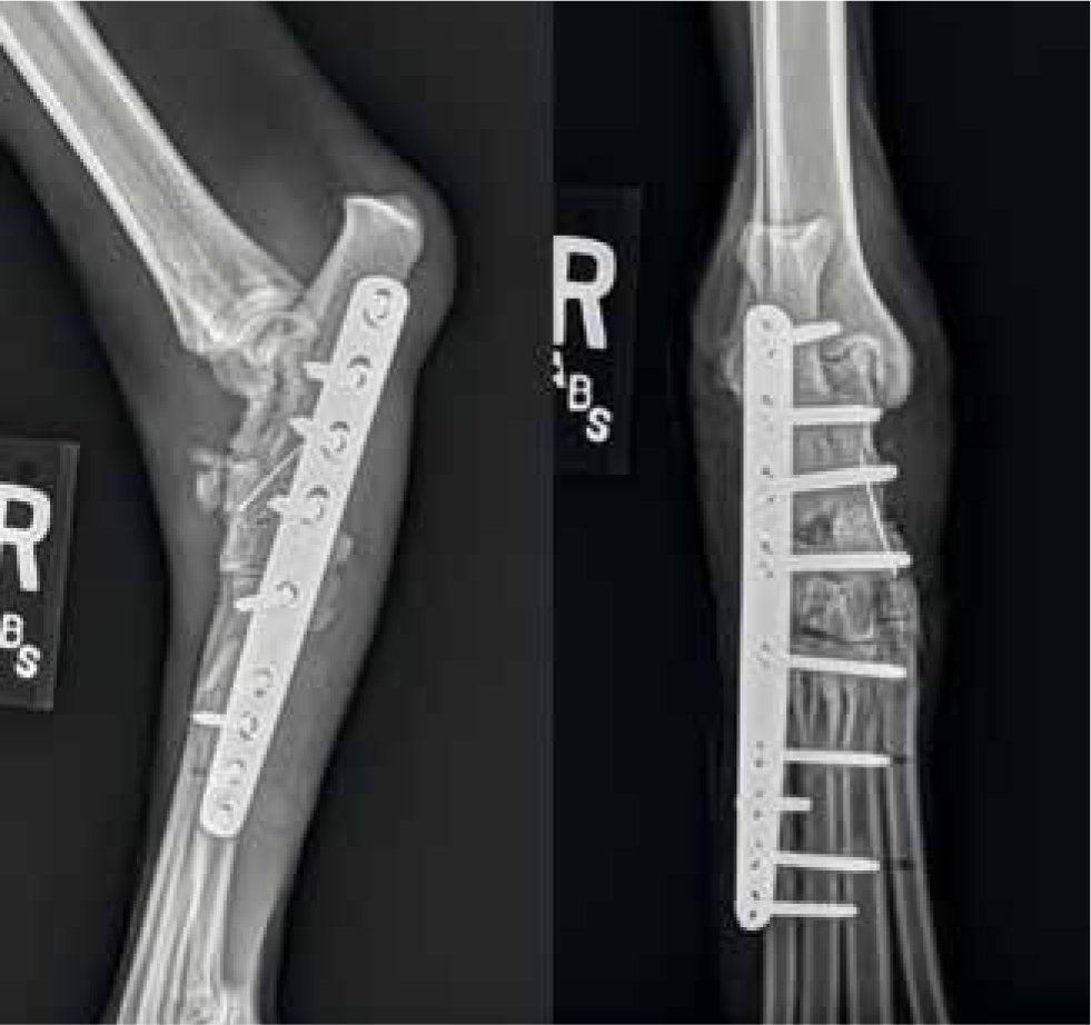
Surgical repair of fractures of the central tarsal bone can be technically difficult. The small size of the dorsal and medial fragments often allows only one chance for the accurate placement of a screw, while the anchor bone itself may be fractured so that the screw has little holding strength (Guilliard, 2000). It is difficult to achieve and maintain tarsal alignment without a well-placed medial lag screw (Guilliard, 2000). In cases where the fragments are too small to allow fixation (Figure 6), including many (but not all) type V fractures, partial tarsal arthrodesis may become the treatment of choice (Figure 7).

Fractures of the plantar process with central tarsal bone luxation generally present with an acute-onset severe pelvic limb lameness, pain on tarsal manipulation and a bony mass palpable on the medial aspect of the tarsus. Mild degrees of plantar convexity and tarsal varus may also be observed (Guilliard, 2007). These dogs may also have concomitant fractures affecting either the calcaneus or fourth tarsal bone, so preoperative images should be carefully reviewed. Surgical repair of these fractures is undertaken through a dorsomedial incision, with the central tarsal bone being reduced and fixed to the fourth tarsal bone with a lag screw; postoperative external coaptation is recommended. Closed reduction and minimally invasive stabilisation of a central tarsal bone luxation has also been reported using a position screw to fix the central tarsal bone to the fourth tarsal bone, with a good outcome recorded at 34 months postoperatively (Hudson and Pozzi, 2012).
Surgical management of fractures of the central tarsal bone results in varying degrees of ankylosis of the intertarsal joints (Boudrieau et al, 1984b), most frequently between the central bone and the fourth tarsal bone, and less commonly between the other tarsal bones. In many cases followed radiographically, total intertarsal ankylosis occurs, with evidence of bridging callus (Guilliard, 2000). Ankylosis even occurs in cases where apparently good interfragmentary reconstruction has been achieved and is thought, in these cases, to be due to the presence of the multiple bone fragments discussed previously that may not be apparent radiographically (Guilliard, 2000). This ankylosis does not appear to be detrimental to the outcome. Some authors have in fact encouraged consideration of removal of the articular cartilage of the intertarsal joints during the primary repair, so as to create an arthrodesis (Guilliard, 2000).
Many Greyhounds are euthanased or retired after sustaining a fracture of the central tarsal bone, although the prognosis for a return to competitive racing after surgical fixation is good (Boudrieau et al, 1984b). The main reasons are the financial cost of the repair; an unacceptably long rest and rehabilitation period; and an assumption that the dog will not regain its previous racing performance (Guilliard, 2000). In fact, in a review of 81 dogs with type III and type IV fractures of the central tarsal bone treated by open reduction and internal fixation, 88% returned to successful racing (Boudrieau et al, 1984b). It is suggested that dogs can be put back into training after 3 months (Bateman, 1958; Houlton, 1998), but radiographs show that ankylosis is not complete at this stage (Guilliard, 2000). The prognosis following reduction and stabilisation of central tarsal bone luxation also appears to be good. In one case series of six Border Collies, all dogs eventually recovered well with normal function being reported, although two dogs did require implant removal in order to resolve persistent lameness (Guilliard, 2007).
Similarly to the literature evaluating calcaneal fractures, the majority of the literature available regarding central tarsal bone fracture/luxation concentrates on the racing dog population and this data cannot necessarily be extrapolated to non-racing dogs. A recent study described traumatic injuries involving the central tarsal bone in nonracing dogs (Armstrong et al, 2019). In contrast to in the racing Greyhound population, the most common diagnosis in this population of nonracing dogs was of a type V central tarsal bone fracture, followed by central tarsal bone luxation. Type III and IV fractures were uncommon in this population. Concomitant fractures of other tarsal bones were common. This altered fracture configuration relative to racing dogs is most likely secondary to the altered aetiology as was discussed for calcaneal fractures; instead of being fatigue fractures, in non-racing dogs the majority of these fractures are secondary to traumatic events. Most of the fractures in the report by Armstrong et al, (2019) were managed surgically either by partial tarsal arthrodesis or using lag or positional screws following open fracture reduction. For dogs with isolated central tarsal bone fractures the prognosis was good following treatment, but the presence of concomitant fractures of other tarsal bones was associated with a higher risk of complications. The differences between racing and non-racing dogs should be taken into consideration when planning treatment and during client communication.
Isolated fractures of the numbered tarsal bones
Second tarsal bone fracture luxations are uncommonly encountered and generally occur along with central tarsal bone fractures. If encountered as an isolated injury, treatment via lag screw fixation to the third tarsal bone is recommended (Carmichael and Marshall, 2012).
Third tarsal bone fractures are reported in isolation but the incidence is low (Prole, 1976; Agnew, 1992). Some cases have a concomitant fracture or luxation of the second tarsal bone (Dee et al, 1990; Guilliard, 2010). Clinical signs are mild, with localised slight swelling and a moderate degree of lameness that rapidly improves; unless radiographs of the tarsus are taken, a misdiagnosis of a sprain may be made (Vaughan, 1987). With time, there may be little local evidence of an injury excepting slight fibrous thickening over the third tarsal bone (Vaughan, 1987). Veterinary examination of Greyhounds with third tarsal bone fractures is often not sought at the time of initial injury, due to the benign presenting signs. Recurrence of lameness after rest is common, however, and with non-surgical management they carry a fair to poor prognosis for return to racing (Dee et al, 1990). Fragment removal is not recommended as a treatment (Guilliard, 2010). The recommended treatment is via lag screw fixation. The prognosis for a successful return to racing appears to be good following fragment fixation in both acute and chronic cases without dorsal tarsal collapse. In cases with dorsal tarsal collapse, a return to racing is less likely (Guilliard, 2010).
Fourth tarsal bone fractures most commonly accompany central tarsal bone fractures. Management of the central tarsal bone injury generally restores anatomical position and stability of the fourth tarsal bone and allows osseous union to progress (Carmichael and Marshall, 2012).
Malleolar fractures
Fractures of the medial or lateral malleoli are well recognised in veterinary practice (Johnson and Boone, 1993; Ness et al, 1996). Fractures may be unimalleolar or bimalleolar and may be associated with shearing injuries (Roch et al, 2009). The medial and lateral malleoli are the anatomical sites for the origin of the medial and lateral collateral ligaments of the tarsus. The arrangement of the malleoli and distal tibia produces a mortise that articulates with the talus (Evans, 1993; Johnson and Boone, 1993). Therefore, with fractures of either the medial or lateral malleolus, talocrural joint instability commonly becomes evident and there may be an apparent luxation (Vaughan, 1987). Clinical signs include swelling, pain and bruising at the distal end of either the tibia or fibula. Instability is demonstrated by applying valgus/varus stress so as to open the joint on the injured side (Vaughan, 1987).
Malleolar fractures are frequently articular in nature; accurate fracture reduction and rigid fixation are desirable to improve joint stability and re-establish the weight-bearing articular surfaces (Boone et al, 1986; Piermattei et al, 2006). This may slow the progression of osteoarthritis and improve joint function in the long-term. Methods for stabilising malleolar fractures include the use of pins, with or without tension band wires, lag or positional screws and a transarticular external skeletal fixator (ESF) (Johnson and Boone, 1993; Owen, 2000; Piermattei et al, 2006). In cats, poor healing appears to be more likely in cases treated with K-wires alone rather than K-wires and a tension band wire (Roch et al, 2009). This indicates that accurate reduction of the fracture and rigid internal fixation of the malleolar fragments with K-wires and a figure-of-8 tension band wire is preferable in this species (Roch et al, 2009). Additionally in cats, malleolar fractures usually involve small articular or juxta-articular fragments that limit the selection of implants, make inadvertent penetration of the joint by the implants more likely and make it difficult to reduce the fragments accurately (Roch et al, 2009).
External coaptation is often recommended postoperatively with either transarticular ESF, dressings or splints. This is commonly left in place for 4–6 weeks, but the optimal duration of coaptation after a malleolar fracture is not known. Perceived advantages of a transarticular ESF over a dressing or splint include better tolerance by the patient (potentially particularly true in cats); better resistance to disruptive forces; fewer complications than after the prolonged application of a splint or cast; and the ability to treat concurrent skin wounds. Disadvantages include longer operations, higher costs, the need for anaesthesia or sedation to remove the frame and the potential for fractures through the pin tracts (Seguin et al, 1997; Owen, 2000; Roch et al, 2009). Hinged transarticular ESF offer potential advantages in maintaining joint mobility and reducing the adverse effects of joint immobilisation on articular cartilage while providing collateral stability (Jaeger et al, 2005). However, hinged constructs are more difficult to apply than rigid frames and are more susceptible to implant-related complications (Jaeger et al, 2005).
Uncomplicated healing is reported in up to 84% of dogs with malleolar fractures treated with open reduction and internal fixation (Boone et al, 1986). In a case series of malleolar fractures in cats (Roch et al, 2009), healing was classified as good or reasonable in 79% of cases; it is worth noting that the small size of the fragments in cats with malleolar fractures can complicate radiographic assessment of fracture healing, owing to the superimposition of metallic implants over the short fracture lines (Roch et al, 2009). Cases that suffer talocrural luxation and significant soft tissue injury may be more likely to suffer a poorer outcome (Schmokel et al, 1994; Owen, 2000), while early postoperative weight-bearing and mobilisation of the joint may improve limb function following surgical treatment (Schmokel and Ehrismann, 2001). Complications encountered following malleolar fracture treatment include non-union, tarsal joint ankylosis, persistent talocrural instability, soft tissue necrosis, implant migration and external fixator pin breakage (Boone et al, 1986; Roch et al, 2009).
Conclusions
Tarsal fractures are common in working breeds but are seen less commonly in the pet population. Fractures in racing Greyhounds are often fatigue fractures or occur secondary to fatigue fractures of the central tarsal bone, while calcaneal fractures in cats may be stress-fractures or associated with KaTS. Achieving an accurate diagnosis is imperative as the first step in making a treatment plan and determining a prognosis; taking multiple radiographic views may assist in the evaluation of this compound joint but computed tomography offers increased sensitivity and facilitates visualisation of all fracture lines as well as appreciation of small fragments which may otherwise be missed. The gold-standard treatment for many tarsal fractures is surgical stabilisation. However, the small fragment sizes and, often, concomitant fractures, render surgery in this area difficult. Additionally, as many tarsal fractures are articular, surgery that does not result in accurate reconstruction and rigid stabilisation may have significant impacts on long-term limb function. Depending on the severity of the injury and other factors, such as owners' financial constraints and willingness to consider the possibility of multiple surgical procedures, joint preservation may not always be a realistic goal. If the joint cannot be preserved, then either pan- or partial tarsal arthrodesis may become appropriate.
KEY POINTS
- Non-racing dogs are more likely to suffer comminuted fractures than racing Greyhounds and therefore the prognosis for future limb function may be more guarded in these patients following tarsal fracture.
- Feline calcaneal fractures may be associated with Knees and Teeth Syndrome and therefore screening for additional fractures and dental anomalies may be warranted.
- Both calcaneal fractures and central tarsal bone fractures are often associated with fractures of other tarsal bones necessitating careful interpretation of diagnostic imaging studies.
- Depending on the severity of the fracture, joint preservation may not be a realistic goal; in these cases arthrodesis may need to be considered.