


This review focuses on some of the new information that has been published around cutaneous adverse food reactions in dogs and cats over the last few years, and considers parallels within the human field that may help guide further work in the prevention, diagnosis and therapy of the disease in dogs and cats.
Food allergy prevention
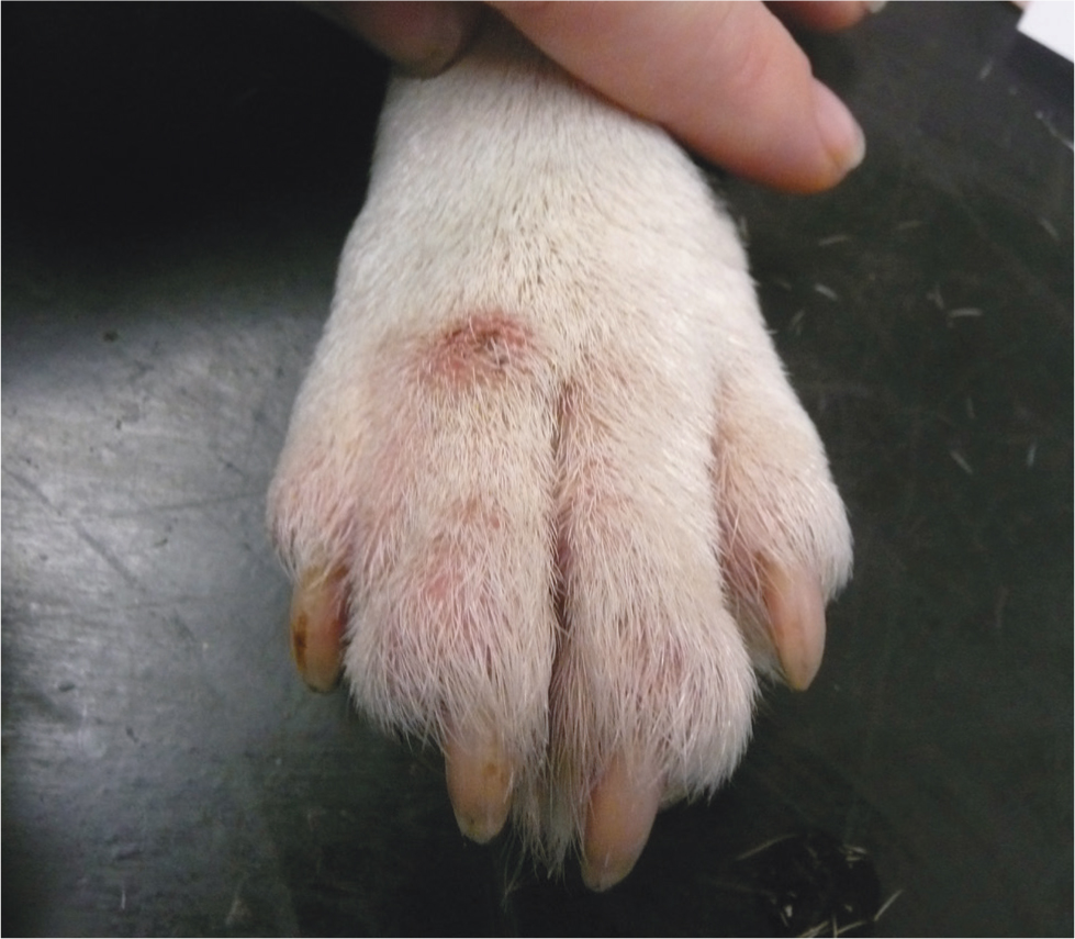
The true prevalence of cutaneous adverse food reactions in dogs and cats is unknown. A critically appraised review of more than 30 articles describing these reactions in dogs and cats found that, among animals presenting with any disease to their veterinary surgeon, the prevalence of adverse food reactions was 1–2% and for those presenting specifically with skin disease, it ranged from 0–24%. Figures increased to 9–40% in dogs with pruritus (Figure 1) and to 8–62% in those with allergic skin disease. In cats with pruritus the range was 12-21%, and 5–13% for those with allergy (Olivry and Mueller, 2016). The authors concluded that cutaneous adverse food reactions should be considered in any companion animal presenting with non-seasonal pruritus or signs of allergic dermatitis. With a relatively high prevalence of disease, an attractive proposition may be to consider how these reactions could be prevented in companion animals. In humans, a range of different interventions have been evaluated as means of preventing ‘food allergy’. These include breastfeeding and the early introduction of allergenic foods to infants at risk for developing food allergy. Current recommendations suggest an emphasis on dietary diversity to include fruit, vegetables and fish during pregnancy, lactation and in early life for infants (Baker and Nowak-Wegrzyn, 2020). Two studies in animals suggested that keeping a dog at home before and during pregnancy decreased the risk of food allergy in 1-year-old children, but the same was not found for other pets such as cats, hamsters, guinea pigs or rabbits (Lyons et al, 2020; Smejda et al, 2020). Whether this is directly related to the presence of the dog or a result of other environmental, dietary or lifestyle choices associated with dog ownership is yet to be established. Similar studies have not yet been undertaken in the veterinary field, although a recent study from Finland suggested a link between feeding pattern and environmental factors in the development of canine atopic dermatitis. A retrospective analysis of 406 dogs with atopic dermatitis suggested that the feeding of a non-processed meat-based diet during the prenatal and early postnatal periods had a significant effect on reducing the incidence of canine atopic dermatitis in adult dogs, compared to the feeding of an ultra-processed carbohydrate-based diet, which increased the risk (Hemida et al, 2020). Other factors that were found to decrease the risk included maternal deworming programmes and sunlight exposure during the early postnatal period (Hemida et al, 2020). On the basis of this retrospective study, it is worth speculating whether the feeding of specific diets during the perinatal period could reduce the incidence of cutaneous adverse food reactions in dogs and cats.
Diagnostic testing in cutaneous adverse food reactions
Numerous papers over the last 10 years have described the use of in vitro tests to diagnose cutaneous adverse food reactions (Jeffers et al, 1991; Jackson, 2001; Bethlehem et al, 2012; Hardy et al, 2014), but none have shown high enough levels of sensitivity and specificity to be a useful substitute for a properly designed exclusion diet. More recent studies have considered again the use of in vitro techniques to diagnose cutaneous adverse food reactions. A German study by Johansen et al (2017) investigated the use of patch testing using different proteins, carbohydrates and dry commercial dog food in dogs with proven cutaneous adverse food reactions, to determine whether the technique had any value in aiding a diagnosis of the condition. The results confirmed that patch testing may be useful for the selection of a suitable protein source for an exclusion diet, but not as a diagnostic tool. Results for raw proteins were found to be more reliable than those for carbohydrates. Patch testing with commercial dog food did not seem to be useful (Johansen et al, 2017). Two other studies have considered serum and saliva-based assays as a means of diagnosing cutaneous adverse food reactions. In the first American study by Lam et al (2019), a cohort of 30 clinically normal dogs were assessed by measurement of food allergen-specific IgE in serum and food-allergen specific IgA and IgM in saliva for 24 different foods. All dogs had a positive test result for at least one assay, suggesting these assays produce positive reactions for normal dogs, making them unsuitable as a diagnostic test. In the second study by Udraite Vovk et al (2019), similar parameters were measured but three groups of dogs were assessed. One group with proven but well-controlled cutaneous adverse food reactions, a second group with allergic dermatitis just starting an elimination diet and a group of clinically normal dogs. No clear difference was found in the positive reactions between the allergic and the healthy dogs, suggesting these tests are not suitable for diagnosis of cutaneous adverse food reactions and elimination diets remain the gold standard for diagnosis. These findings therefore suggest that while in vitro tests may be useful to help select the ingredients for an exclusion diet, an exclusion diet should remain the diagnostic test of choice.
Selection of novel proteins and cross-reactions of food
While the institution of a hypoallergenic diet is key to making a diagnosis of cutaneous adverse food reaction, the selection of the components of that diet can be difficult. Several factors contribute to this:
- Selecting a novel protein that the dog has not had contact with before and that does not cross-react with other components of their diet
- Ensuring that there are no undeclared foods in a commercially prepared diet.
It has been recognised for some time in human medicine that cross-reactivity occurs between different food allergens. For example, individuals who are allergic to milk may also be allergic to beef and pork (Mamikoglu, 2005), or those allergic to chicken may also be allergic to fish (Kuehn et al, 2016). Cross-reactivity also occurs between seeds and cereals (Hemmer et al, 2017). Many of the foods that show marked signs of cross-reactivity in humans form some of the most common foods to cause cutaneous adverse food reactions in dogs, namely beef, dairy products, chicken and wheat (Mueller et al, 2016). Animal studies have suggested that cross-reactivity may occur between beef, lamb and cow's milk (Bexley et al, 2017), as well as between fish and chicken (Bexley et al, 2019), suggesting that closely related protein or carbohydrate sources should be avoided when formulating a novel protein diet. While there is some reluctance among dog owners to feeding their pets a wholly vegetarian/vegan diet, because of the perception that such diets are unbalanced (Dodd et al, 2019), other diets containing mixtures of amino acids (Kawarai et al, 2010) or insects (Bohm et al, 2018) have been successfully used as novel exclusion diets. The use of hydrolysed diets negates the need to search for a novel food source. Hydrolysis of the proteins within these foods disrupts the proteins, rendering them hypoallergenic. While potential problems with these diets include a reduction in palatability and a risk of diarrhoea, there is a wealth of evidence to show they are effective (Cave, 2006; Bizikova and Olivry, 2016; Matricoti and Noli, 2018). In one study, extensive protein hydrolysation of the poultry feather extract of a diet prevented the allergenic epitopes being recognised by poultry-specific IgE (Olivry et al, 2017).
Several studies have identified another major problem with the use of commercial rather than home-cooked exclusion diets, which is the presence of undeclared proteins (Ricci et al, 2013; Horvath-Ungerboeck et al, 2017; Kanakubo et al, 2017; Fossati et al, 2019). This has been identified in both meat- and vegetable-based exclusion diets (Kanakubo et al, 2017). In many cases, proteins have been identified that are not noted within the food labelling and in some cases, diets have been found not to contain the proteins listed on the label. Without a complete knowledge of the components of a diet it is impossible to assess a response to therapy. Of course, there are exclusion diets that are not contaminated and hydrolysed diets appear to be relatively less affected than other diets (Horvath-Ungerboeck et al, 2017).
Based on existing knowledge of the field, the current recommendation for designing a hypoallergenic diet would be that a home-cooked diet is still ideal, providing a truly novel food source can be used. Alternatively, a highly hydrolysed commercial exclusion diet should be used in a food trial for a dog to identify cutaneous adverse food reactions.
Storage mites in dog food and implication for food trials
House dust mites of the genus Dermatophagoides are recognised as the most common inciting allergen, as recognised by circulating IgE levels in dogs with atopic dermatitis (Nuttall et al, 2006). Several studies have described cross-reactivity between house dust mites (Dermatophagoides pteronyssinus and Dermatophagoides farinae) and storage (forage) mites Acarus siro, Tyrophagus putrescentiae and Lepidoglyphus destructor. In addition, IgE reactivity against storage mites is common in dogs with atopic dermatitis (Arlian et al, 2003; Saridomichelakis et al, 2008; Buckley et al, 2013). Cross-reactivity is probably clinically relevant, as beagles experimentally sensitised to D.farinae exhibit flares of their clinical signs when challenged environmentally or orally with the storage mite Tyrophagus putrescentiae (Marsella and Saridomichelakis, 2010). Storage mites are known to occur in dry dog food and in one study, paper bags were shown to create an environment conducive to mite multiplication compared to food stored in plastic bags or boxes (Gill et al, 2011). Where kibble is stored in conditions of high relative humidity and becomes contaminated with mould, mite viability is increased (Canfield and Wrenn, 2010). A Spanish study showed that under low-to-average temperatures (16°C) and humidity (68%), mites were undetectable in bags of dog food over a 6-week period; whereas food stored at higher temperature (average of 23°C) and humidity (average 72%) became contaminated, with 80% of opened bags and 67% of unopened bags being found to harbour Tyrophagus putrescentiae mites (Brazis et al, 2008). Although this was a Spanish study, there is no doubt these types of environmental conditions could be replicated in an indoor environment, such as a kitchen, where dog food may be stored. The important take home message from these studies is that despite the high efficacy of hydrolysed diets in diagnosing cutaneous adverse food reactions, if dried forms of these diets are stored poorly and contain storage mites it is possible that house dust mite allergic dogs eating the food could have an allergic flare, leading to a false diagnosis. When commercial dried dog food is used to diagnose cutaneous adverse food reactions, especially to feed dogs with a house dust mite allergy, they should be newly purchased and stored appropriately. Ideally, foods should be stored in plastic boxes in low-to-average temperatures and humidity to prevent a false diagnosis and reduce the likelihood of flares in dogs that are allergic to house dust mites (Olivry and Mueller, 2019).
Time to flare of cutaneous signs after a dietary challenge
The final article in the evidence-based series of critically appraised topics on adverse food reactions, considers the time to flare-up of cutaneous signs in dogs and cats with food allergies after dietary challenge (Olivry and Mueller, 2020). While laboratory and in vivo tests cannot be used reliably to diagnose cutaneous adverse food reactions in dogs and cats, the institution of an appropriately formulated exclusion diet remains the gold standard. However, the institution of an exclusion diet and a significant improvement in clinical signs are not enough to establish a diagnosis. The seasonal nature and spontaneous improvement of atopic dermatitis (which can occur concurrently to a cutaneous adverse food reactions) can lead to a misdiagnosis of a food reaction.
It is advisory to undertake a provocative challenge with ingredients of the previous diet to establish the existence of a cutaneous adverse food reaction and identify the offending allergens. Dietary challenge is usually labour intensive and owner compliance can be poor if they have had to feed their pet on a restricted diet for a considerable period. Therefore, the knowledge of the shortest time that an ingredient needs to be fed in order to provoke a reaction is important. A review of nine papers that included 234 dogs and four papers that included 83 cats were evaluated for evidence (Olivry and Mueller, 2020). Few animals (9% dogs, 27% cats) flared on the first day after challenge, which suggests that an IgE mediated pathogenesis may only be responsible for a small number of cases. At 7 days post-challenge, 80% of both dogs and cats had flared and a 90% flare rate was observed in dogs at 14 days and cats at 7 days. It may be that the small numbers of papers reviewed, together with the underreporting of clinical observations in the first few days after challenge, may have skewed these results. However, the data suggest that a flare should be seen in most animals within a week of introducing the original diet and that if individual ingredients are to be assessed, they should probably be added every 7 days.
Sublingual therapy for cutaneous adverse food reactions
Food allergies can have dramatic and life-threatening consequences in humans. As a result, a huge amount of work has been undertaken to evaluate the potential benefits of immunotherapy as a means of desensitising people to specific food allergens. Food allergen-specific sublingual immunotherapy (FA-SLIT) and oral immunotherapy (FA-OIT) in humans have shown promise as therapeutic interventions in individuals who are allergic to peanuts, cow's milk, egg, wheat and many other foods (Nowak-Wegrzyn et al, 2019). While the results obtained from the use of FA-OIT are superior to FA-SLIT, the safety of FA-OIT is less favourable. Ongoing work in humans will be aimed at optimising and standardising protocols for administration and maximising their safety (Nowak-Wegrzyn et al, 2019; Albuhairi and Rachid, 2020; Waldron and Kim, 2020). As a result of this work in the human field, several studies have been undertaken to assess the potential benefits of FA-SLIT in dogs. An initial study to investigate the safety, tolerability and dispenser sterility of FA-SLIT in healthy dogs, before testing it in food allergic dogs, showed the therapy was well tolerated and safe (Maina et al, 2016). A further double-blind, randomised, placebo-controlled trial of FA-SLIT in client-owned dogs with adverse food reactions by the same investigators showed that the therapy was effective, well tolerated and safe. Although only small numbers of dogs were included in this pilot study, the treatment was rated as effective or quite effective by 80% of owners, and the placebo of glycerinated saline was rated as ineffective by all owners. Statistical tests showed significant protection against food challenge-induced clinical signs as measured by pruritus visual analog scale and canine atopic dermatitis extent and severity index scores. Two dogs were withdrawn from the study because of an exacerbation of their clinical signs by FA-SLIT (Maina and Cox, 2016). Further study investigating changes in key cytokines associated with FA-SLIT showed significant differences between the treatment and placebo groups, with increases in Interleukin-10 and Interferon-gamma in the treatment group compared to the placebo group. These findings suggest FA-SLIT may modulate the immune response towards a T-helper type 1 and regulatory T cell phenotype to create tolerance in dogs with cutaneous adverse food reactions (Maina et al, 2017) and may offer an option for treatment in dogs.
Summary
While there is no hard evidence to support the use of an exclusion diet to prevent the development of food allergy in dogs and cats, based on the results of studies in humans, it is possible that strategies may be developed in the future. Despite the availability of new diagnostic tests, the institution of a home-cooked diet containing a novel protein remains the gold standard test for diagnosing cutaneous adverse food reactions. In vitro tests may be useful to help select the components of the diet. However, clinicians need to be aware of the risk of cross-reactivity of different foods and the potential for contamination of commercial diets with undeclared proteins, or with forage mites if they are not stored properly. A definitive diagnosis of a cutaneous adverse food reaction should involve challenge with the original diet and its components after a clinical resolution of signs has been achieved. The introduction of each new food should be done at weekly intervals so that the response to the addition of each new food can be fully assessed. FA-SLIT is a promising therapeutic option for the management of cutaneous adverse food reactions, but further work in larger cohorts of dogs to assess the long-term benefits is needed.
KEY POINTS
- In vitro tests may be useful to help select ingredients for an exclusion diet, but an exclusion diet should remain the diagnostic test of choice for cutaneous adverse food reactions.
- An ideal exclusion diet should be home cooked, providing a truly novel food source can be used that does not cross-react with other foods, but a highly hydrolysed exclusion diet can offer a good alternative if this cannot be formulated.
- Commercial diets can be contaminated with undeclared protein sources, making interpretation of results difficult.
- Dried diets should be stored in plastic boxes, in conditions of low average temperature and humidity, to prevent contamination with forage mites as this can lead to a false diagnosis of cutaneous adverse food reactions.
- When dietary challenge is performed to confirm a diagnosis of cutaneous adverse food reaction, most animals will flare within a week of introducing the original diet.
- Sublingual therapy may provide an option for therapy for cutaneous adverse food reactions in dogs and cats in the future