Locoregional anaesthesia of the pudendal nerves and their branches has been described in a variety of species for surgical procedures of the genitalia (Edwards, 2001; Perkins et al, 2003; Adami et al, 2014; Fazili et al, 2016; Gallacher et al, 2016). The pudendal nerves arise bilaterally from the ventral branches of the three sacral nerves that constitute the sacral plexus (Bailey et al, 1988). The left and right pudendal nerves exit the pelvis caudally at the pelvic outlet. In males, these nerves continue as the left and right dorsal nerves of the penis, which course caudally over the dorsal aspect of the ischiatic symphysis either side of the midline urethra (Evans and de Lahunta, 2013). Each dorsal nerve of the penis is closely associated with the corresponding penile artery and vein, together providing the main sensory afferent innervation of the penis (Evans and de Lahunta, 2013). The caudal rectal and perineal nerves branch off the pudendal nerves more proximally at the level of the pelvic outlet, providing sensory and motor innervation to the external anal sphincter, perineum and proximal penis (Evans and de Lahunta, 2013).
In cattle, the pudendal nerve is commonly approached by advancing a needle via the ischiorectal fossa into the sacrotuberous ligament, guided by palpation per rectum (Edwards, 2001). This approach is referred to as an internal pudendal block (Edwards, 2001). Gallacher et al (2016) demonstrated the provision of anaesthesia in horses requiring genitourinary surgical procedures using a similar needle entry point at the ischiorectal fossa, with electrolocation to identify the pudendal nerve. In goats and horses, anaesthesia of the branches of the pudendal nerve has been successfully performed at the level of the ischial arch, circumventing the need for palpation per rectum (Perkins et al, 2003; Fazili et al, 2016) and targeting the dorsal nerves of the penis for penile surgical procedures. Ultrasound-guided locoregional approaches to the pudendal nerve have also been described and investigated in cats (Adami et al, 2013) and donkeys (El-Khamary et al, 2017). In cats, this approach was successful in providing peri-operative analgesia for perineal urethrostomy (Adami et al, 2014). In donkeys, the internal pudendal vessels were used as anatomical landmarks for the location of the pudendal nerve, which was not visualised during ultrasonography (El-Khamary et al, 2017). The success of this technique was confirmed by loss of sensation in the penis in all donkeys and dissection to reveal dye spread over the pudendal nerve (El-Khamary et al, 2017).
As far as the authors are aware, locoregional blockade of the dorsal nerves of the penis in dogs for penile urethral surgery has not been described, although it represents a targeted locoregional approach to complement a multimodal analgesia protocol in suitable patients.
Presentation
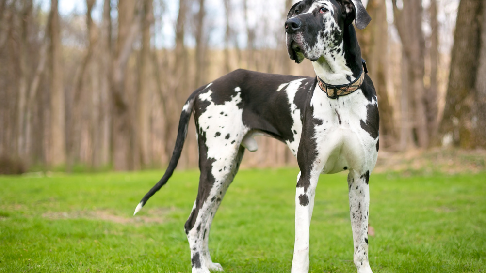
A 56.7 kg male neutered Great Dane, aged 3 years and 5 months was referred with a 4-month history of recurrent urethral prolapse and haematuria, refractory to purse string placement under general anaesthesia at the referring veterinary practice.
On presentation, the dog was alert but anxious and panting. Heart rate was 120 beats per minute (bpm) with synchronous, strong peripheral pulse quality, and thoracic auscultation was unremarkable. Mucous membranes were pink and moist and capillary refill time was < 1.5 seconds. A body condition score of 5/9 was assigned (Laflamme, 1997). The urethra had prolapsed 5 mm at the external urethral meatus, with a swollen and red appearance, which was visible on extrusion of the penis. Pain scoring was not possible because of the location of the abnormality and the dog's anxious demeanour. Visual evidence of inflammation of the prolapsed urethral mucosa indicated this was likely to be painful for the dog. The client consented to anaesthesia and its associated risks, as well as diagnostic imaging and surgical management.
Ultrasonography of the urinary tract was otherwise unremarkable. The dog was assigned an American Society of Anesthesiologist physical status classification of 2 (Portier and Ida, 2018) and scheduled to have a distal urethral resection and anastomosis under general anaesthesia.
Management
A 20G intravenous (IV) cannula (Jelco; SurgiVet) was placed aseptically in the left cephalic vein and the dog was premedicated with methadone (0.2mg/kg) (Methadyne; Jurox) and dexmedetomidine (2 µg/kg) (Dexdomitor; Zoetis) IV. Following pre-medication, after 5 minutes the dog laid down quietly and became unresponsive to noise stimulation. Anaesthesia was induced with propofol (Propoflo; Abbott), titrated to effect over 120 seconds (2.6 mg/kg) until an adequate plane of anaesthesia allowed tracheal intubation with a size 13 mm, cuffed, polyvinylchloride endotracheal tube (AirCare; Smiths Medical ASD Inc). The endotracheal tube was connected to a circle breathing system (Eickemeyer) and sevoflurane (SevoFlow; Zoetis) (2-3%) was delivered in oxygen (4 L/min for first 15 minutes, reduced to 1.5 L/min thereafter) to maintain anaesthesia. Multiparametric monitoring was performed every 5 minutes and included oxygen saturation, capnography, electrocardiography, oscillometric blood pressure, temperature, gas monitoring and spirometry (Datex Ohmeda S5; GE Healthcare). The plane of anaesthesia was monitored every 5 minutes using jaw tone, eye position and palpebral reflex assessment. Hartmann's lactated ringers (B. Braun) was administered at 4 ml/kg/hour during surgery. Cefuroxime (20 mg/kg) (Zinacef; GlaxoSmithKline) was administered once via IV after instrumentation.
The patient was positioned in right lateral recumbency and the perineum clipped and aseptically prepared with chlorhexidine and alcohol. An assistant extended the tail and an 8.5 MHz linear ultrasound transducer (UGEO PT60A; Samsung) was placed on the perineal region with its long axis at right angles to the dorso-ventral midline (Figure 1). The ischial arch was visible as a hyperechoic elongated structure, along with the paired left and right vessels of the penis, either side of the urethra (Figure 2). The presence of blood flow within the identified vessels was confirmed by colour flow Doppler (Figure 3).
Two hypoechoic structures surrounded by hyperechoic tissues, located next to the identified vessels, were visible symmetrically either side of the urethra. No Doppler flow was present within these structures, and they were assumed to be the intended target nerves for the regional nerve block. This location was consistent with the reported location of the dorsal nerves of the penis (Evans and de Lahunta, 2013). A 22G, 50 mm insulated needle (Stimuplex Ultra 306, B. Braun) was inserted in-plane, approaching the left and right identified structures in turn. Aspiration prior to each injection revealed no blood and 5 ml of 0.5% weight by volume bupivacaine (B. Braun) was infiltrated (0.18 ml/kg; 0.9 mg/kg total dose). Injection was subjectively assessed as having little to no resistance and the injectate was visualised to surround the identified structures on the ultrasound image.
The dog was transferred to theatre and positioned in dorsal recumbency. Surgical resection of the prolapsed urethra and anastomosis of the healthy tissue commenced 35 minutes after the nerve block. During the surgical procedure, HR remained between 110-120 bpm, respiratory rate between 8–9 bpm and mean arterial pressure at 71-78 mmHg. The dog ventilated spontaneously during anaesthesia. The duration of the surgical procedure was 25 minutes and the total anaesthesia time was 80 minutes.
Intravenous fluid therapy was discontinued and the dog was transferred to a recovery room and extubated 10 minutes after disconnection from the breathing circuit. Recovery was smooth without any apparent distress and the dog stood 15 minutes after exubation with no ataxia. No penile protrusion was observed. Postoperative analgesia was provided with meloxicam (0.2 mg/kg IV loading dose, followed by 0.1 mg/kg per os every 24 hours) (Metacam; Boehringer Ingelheim) and buprenorphine (0.02 mg/kg IV every 8 hours for two doses, starting 4 hours after pre-medication) (Buprecare; Animalcare). Postoperative pain scoring was not possible because of the anxious temperament of the dog and the difficulty accessing the surgical site. Given the lack of interference at the surgical site, the dog was assessed to be comfortable postoperatively. Normal urination was observed 3 hours postoperatively.
Outcome
The dog was discharged 24 hours after the surgical procedure with no complications. Unfortunately, recurrence of the urethral prolapse occurred 7 days postoperatively, although the dog displayed no urinary abnormalities or discomfort. The dog is currently being monitored for recurrence of urinary abnormalities that would require repeated surgical intervention.
To confirm that the structures identified using ultrasound in the clinical case were the dorsal nerves of the penis, an ultrasound-guided injection was repeated using a 12kg male canine cadaver with the same method as described, without the use of a colour flow Doppler. The use of the cadaver for teaching and research had been previously consented to by the owner. Methylene blue dye (1% weight by volume) was injected at a volume of 0.18 ml/kg and was seen to disperse around the location of the identified structures believed to be the dorsal vessels and nerves of the penis in a similar manner to the case described. Dissection down onto the injection site revealed bilateral structures considered to be the proximal dorsal nerve, artery and vein of the penis coursing around the ischium in the region of the dye (Figure 4). This finding was consistent with the area that the dorsal nerves of the penis are described to run (Evans and de Lahunta 2013).
Discussion
This case report describes ultrasound-guided local anaesthetic infiltration in the region of the dorsal nerves of the penis via a transperineal approach in a dog. It was possible to visualise the left and right dorsal vessels of the penis, urethra and ischial ridge using ultrasonography to facilitate perineural local anaesthetic deposition. No adverse events relating to the nerve block were observed and the multi-modal analgesia plan was successful in managing pain throughout the surgical and recovery period. It was not, however, possible to be certain that complete anaesthesia of the surgical site was achieved with the local anaesthetic injection. Subsequent dissection performed on a cadaver suggested that the technique described demonstrates the potential to deposit injectate around the location of the intended nerves.
Epidural injection of local anaesthetic represented an alternative technique to provide anaesthesia of the surgical site described in this case report. Perineural infiltration around the dorsal nerves of the penis provides a more targeted approach, without complications relating to epidural administration of local anaesthetics, such as hypotension. Epidural administration of local anaesthetic can also risk impairing the hind limb motor control (Edwards, 2001; Olbrich and Mosing, 2003; Cyna and Middleton, 2008; Grubb and Lobprise, 2020), complicating postoperative nursing care. This was particularly pertinent in this case, given the large body mass and stressed demeanour of the dog. Alternative approaches include local infiltration or direct application (also referred to as a ‘splash block’) of anaesthetic agent at the surgical site. Infiltration would not have been possible in this case because of the risk of distortion of the surgical site. A splash block may have represented a simple alternative approach, but the effectiveness of perioperative analgesia has not been investigated in this anatomical location. Clinical studies investigating the effectiveness of analgesia after splash blocks in differing anatomical locations have reported variable results (Thompson and Johnson, 1991; Winkler et al, 1997; Zibura et al, 2019).
The transperineal approach to the dorsal nerve of the penis at the ischial arch was selected, as only the penis and distal urethra required anaesthesia for the planned procedure. This nerve carries primarily sensory afferent fibres (Evans and de Lahunta, 2013), so nerve stimulation would not have evoked a motor response, ruling out the possibility of a nerve stimulator guided approach. Furthermore, this precludes the assessment of absence of motor tone postoperatively as a marker for ongoing successful nerve blockade. More proximal blockade of the pudendal nerve, with the aid of nerve stimulator guidance, is possible, as branches of the pudendal nerve supply motor innervation to the anus, perineum and retractor penis muscle, which has been described in horses (Gallacher et al, 2016). It confers the additional benefit that assessment of anal tone could be used as an indicator of successful blockade, particularly in the postoperative period.
A disadvantage of the ultrasound-guided technique described in this case report was the inability to determine the success or duration of the block. The surgery was likely to elicit nociception as sensitive tissue resection was performed, but it was possible that the lack of response to surgical stimulation was because of the systemic analgesics administered. It is also possible that the prolonged prolapse of the urethral mucosa and/or previous attempts to suture the prolapse in place may have influenced the sensory innervation to the tissues. The dye deposition observed in the cadaver supports the theory that the technique has potential to provide at least partial anaesthesia to the tissues innervated by the dorsal nerves of the penis. There are limitations associated with interpretation of dye deposition in the cadaver compared to the clinical case in this report. These include the influence of tissue damage secondary to freezing, storage and thawing of the cadaver on the spread of dye.
Possible complications of blockade of the pudendal nerve and its branches include intra-vascular injection, local anaesthetic toxicity or hypersensitivity, nerve or vascular damage, infection, incomplete blockade, and damage to the rectum and/or urethra (Campoy and Schroeder, 2013). Careful technique, good visualisation of structures prior to needle placement and aspiration prior to any injection should minimise the chance of these complications. Incorrect needle placement or extensive local anaesthetic spread also carries the risk of locomotor impairment, as inadvertent blockade of the sciatic and caudal gluteal nerves is possible (Gallacher et al, 2016), although unlikely, during the ischial arch approach. In humans, ultrasound-guided peripheral nerve blocks confer some benefits over nerve stimulator-guided approaches, such as fewer needle insertions (Koscielniak-Nielsen, 2008). It is important to note the benefits of ultrasound-guided approaches are dependent upon and limited by the acquisition of adequate image quality to facilitate identification of important structures (Koscielniak-Nielsen, 2008). Adami et al (2013) encountered this problem on attempting a transperineal approach to the pudendal nerve in cat cadavers. The approach was judged as clinically inappropriate in cats because of the small visual field and high technical difficulty (Adami et al, 2013). The latter could have been a result of the relatively poor spatial resolution achieved with the 7.5 MHz ultrasound probe used (Adami et al, 2013). Musculoskeletal ultrasound takes place at a similar tissue depth, with frequencies more than 10 MHz recommended (Penninck and d'Anjou 2015). El-Khamary et al (2017) also failed to visualise the pudendal nerve trunk in donkeys using an ultrasound-guided ischiorectal fossa approach, but successfully used nearby vasculature as a guide for local anaesthetic deposition. Small ultrasonographic field could pose a technical constraint in smaller dogs but may be negated by using higher frequency ultrasound probes to improve the spatial resolution of key structures. The technique described was also performed in the cadaver, which was 12kg. Ultrasonographic identification of the urethra, ischial arch and blood vessels was subjectively deemed straightforward by the anaesthetist performing the procedure in this case report, although identifying the nerves with unequivocal certainty was more challenging.
An ischial arch approach to local blockade of the pudendal nerve, like that used in this case report, has been described in goats (El-Kammar and Alsafy, 2006). This approach was associated with a reduced incidence of inadvertent rectal or arterial puncture compared to the ischiorectal fossa approach (Fazili et al, 2016). In goats, the dorsal nerves of the penis are reported to provide motor innervation to the retractor penis muscle (El-Kammar and Alsafy, 2006), so the observation of penile protrusion following local anaesthetic administration could indicate successful blockade of the dorsal nerves of the penis. Interestingly, spontaneous protrusion of the penis from the prepuce was not observed by Fazili et al (2016) following either the ischial arch or ischiorectal fossa approach. In dogs, the retractor penis muscle is innervated by the deep perineal branch of the pudendal nerve (Evans and de Lahunta 2013), therefore spontaneous penile protrusion was neither anticipated nor observed following the locoregional approach described in this case report.
Postoperative pain scoring was not used in this case because of the difficulty assessing pain at the surgical site and the stressed demeanour of the patient. Systemic analgesia was administered postoperatively to ensure analgesia was provided in the recovery period. It may not have been necessary to provide additional analgesia this early in the postoperative period if the block was effective. The patient and economic benefits of reduced postoperative systemic analgesics provided by local regional anaesthesia have been previously demonstrated (Palomba et al, 2019).
Further research is required to investigate the success rate of the described technique before it can be widely recommended for distal penile surgery. Further imaging and cadaveric studies are also required to elucidate the suitability of different breeds of dog and any associated complications.
Conclusions
In this case, an ultrasound-guided, transperineal approach for perineural local anaesthetic injection around the bilateral dorsal nerves of the penis, using the urethra and dorsal arteries of the penis as landmarks, is described. Adequate analgesia was achieved with the multimodal plan administered and no adverse events relating to the nerve blockade were encountered.
KEY POINTS
- Ultrasound-guided locoregional blockade of the dorsal nerve of the penis can be considered as part of a multimodal analgesia protocol for surgical procedures of the distal urethra in dogs.
- Locoregional techniques described for large animals may not always be directly translatable to companion animals.
- It is imperative to ensure good visualisation of local anatomical structures during ultrasound guided locoregional blockade.
- Further studies are required to elucidate the success and complication rate of the block described and the suitability of different breed and size of dogs for this approach.