


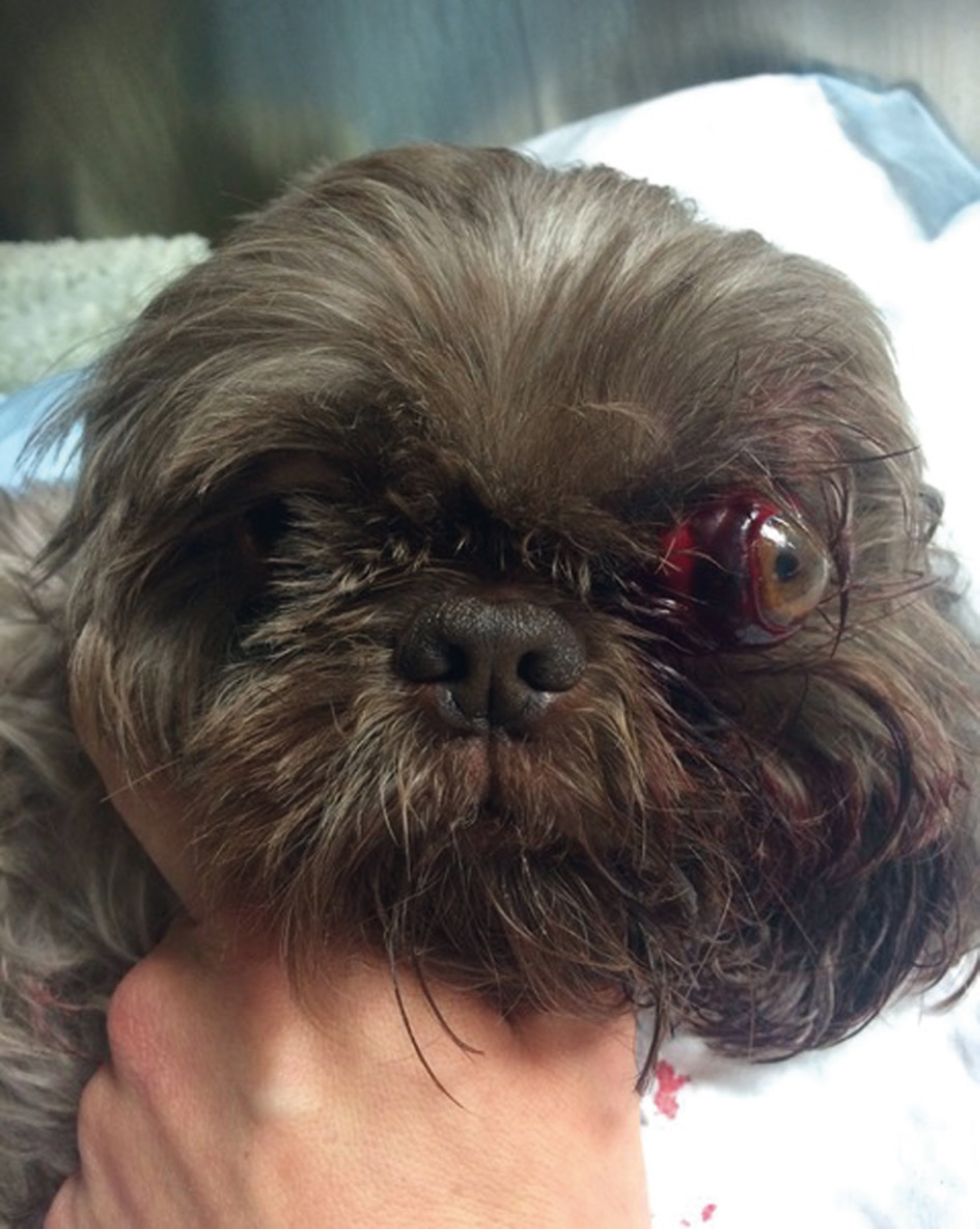
It is a sunny Sunday afternoon, and you have just sat down for lunch. The emergency phone rings, and on the other end of the line is a distressed owner who informs you that their dog Molly, a 2-year-old female Shi-Tzu, has been in a fight with another dog and is now bleeding from the left eye. You agree to meet them at the practice. On presentation Molly is in significant pain, and therefore reluctant to be examined. She is very hairy, and thus complete assessment of the damage if difficult, however the left globe is clearly protruding when compared with the right (Figure 1).
Question 1 — You decide you are going to have to anaesthetise Molly to fully assess her, as she is currently in too much pain to tolerate handling. You have deemed that there are no other life-threatening injuries present, and that it is safe to do so. What parts of the ophthalmic examination are vital to be performed before inducing anaesthesia in this case?
Answer — When assessing proptosis cases there are two distinct questions the clinician should consider following stabilisation of the patient: 1) what is prognosis for vision? and 2) what is the prognosis for the globe? The answer to both questions will allow you to make an appropriate therapeutic plan. Assessment of potential for vision can, and should, be performed at this stage, before anaesthesia, with minimal handling:
- The first test (and offen forgotten) is the menace response. Do not forget that, if the globe is proptosed, the eyelids will be trapped behind the equator of the protruding globe, and therefore a ‘blink’ per se will not be observed. However, these animals will offen pull back their head or try and move away from the menacing hand. A normal menace response is obviously a good prognostic indicator, yet an absent menace response may not necessarily mean all is lost, and you should progress to the next step
- Direct pupillary light reflex (to the affected eye) and/or consensual pupillary light reflex (reflex in the unaffected eye, when stimulating the affected eye) have been shown to be positive prognostic indicators for vision following globe replacement. Remember to use a bright light source for this test (a pen-light may not suffice).
Assessment of the prognosis of the globe itself would require handling, and in this case should be performed once the patient is anaesthetised due to the considerable pain the patient is suffering.
In Molly's case, there was an absent menace response, and no direct or consensual pupillary light reflexes.
Question 2 — You have induced general anaesthesia, what are you looking for at this stage?
Answer — There is a considerable amount of discharge and matted hair, this should all be removed to clean the area, and assess the full extent of the damage. Once this is performed, we can now assess prognosis for the globe.
Distant examination:
- Extra-ocular muscles: avulsion of more than one extra-ocular muscle is associated with a poor prognosis
- Globe rupture: loss of integrity of the globe, either by direct trauma or compressive forces, can lead to long-term pathology, including glaucoma, endophthalmitis and phthisis bulbi. In these cases, if the globe is eventually successfully salvaged, it is important to inform the owner that they may have to administer life-long medication to retain a blind eye
- Damage to peri-ocular structures: depending on the cause of the trauma, concurrent facial fractures or soff tissue damage may be present and should also be addressed.
Close examination:
- The remainder of the ophthalmic examination may now be performed, including assessment of the cornea and intraocular structures, fluorescein testing, and intra-ocular pressure if possible
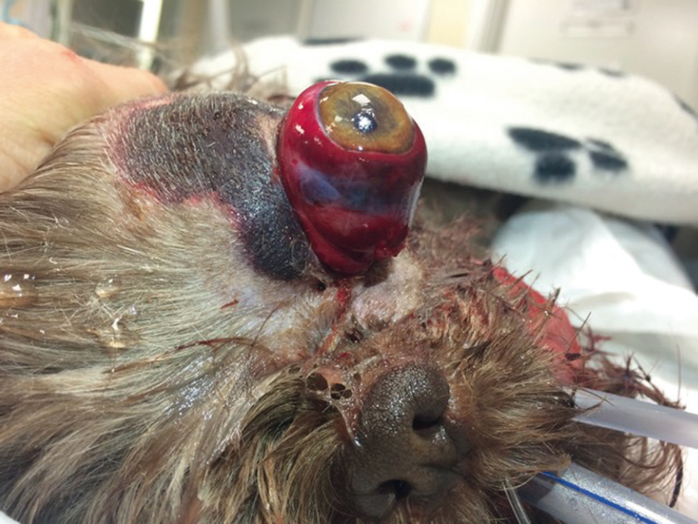
- At this stage, B-mode ocular ultrasonography could be considered, and may provide information regarding the integrity of the globe, and location of intra-ocular structures. In the present case, Molly had avulsion of >3 extra-ocular muscles, leading to severe proptosis and globe deviation, 360o subconjunctival haemorrhage, and marked periocular bruising with no palpable fractures (Figure 2).
Question 3 — Based on your findings what would be your recommendations for this case? What factors would change your decision, and how would that affect the course of action?
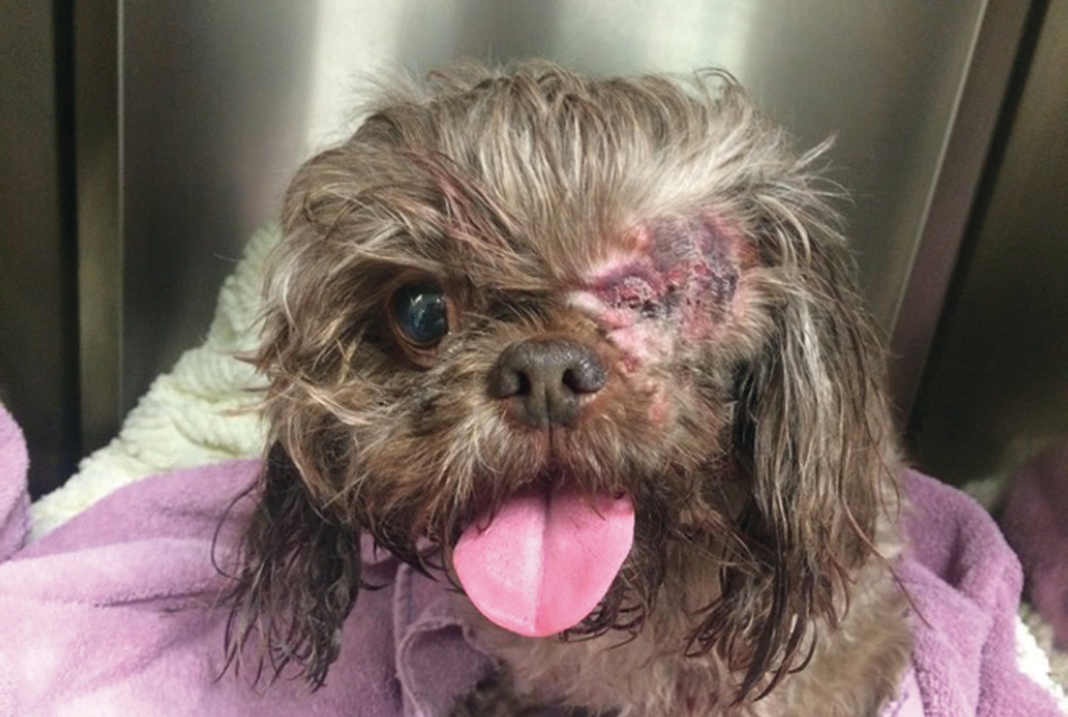
Answer — In the present case, the absent menace response, absent direct and consensual pupillary light reflexes (PLRs) and the avulsion of more than one extra-ocular muscle with marked globe proptosis lead to a very poor prognosis for both vision and globe. As such, enucleation was recommended (Figure 3). However, whenever salvage of the globe is possible this should be attempted (via replacement of the globe and temporary tarsorrhaphy for at least 3 weeks). Situations where replacement may be attempted include:
- If a menace response and/or PLRs are present on admission, then globe replacement would certainly be recommended with an improved prognosis for vision.
- If there is no menace response or PLRs, however the structural damage is minimal, then replacement of the globe could be attempted, if this was the owners choice. The prognosis for vision in this case would be poor, however the globe could still be retained for cosmetic appearance, although strabismus is common after replacement.
Question 4 — What other factors would you consider for Molly's long-term care?
Answer — Following enucleation of the left eye, the ocular health of the remaining right eye is of the utmost importance (Figure 3). Owing to her breed-associated conformational exophthalmia, Molly has a higher risk for potentially globe threatening ocular pathology, such as corneal ulceration. Strategies, such as ongoing lubricants and medial canthoplasty surgery to improve her eyelid conformation, and thus her corneal health, should therefore be discussed with the owner. Additionally, client education on the clinical signs of ocular disease in the remaining eye, and the subsequent urgency of reporting these to a veterinary surgeon as soon as they occur, are essential for the ongoing health of the right eye.
Conclusions
Decision-making process around a traumatic proptosis case:
- Stabilise the patient, ensuring lubrication of the cornea while doing this
- Prioritise the parts of the ophthalmic examination that can be performed on a conscious animal
- Assess for prognosis of both vision and globe, and adjust owner expectations accordingly
- If the globe is salvageable, attempt globe replacement with temporary tarsorrhaphy, vision may or may not return but in general the prognosis for vision in these cases is poor
- If the prognosis for the globe is poor, enucleation may be required.
Please note that this self-assessment has focused on the decision-making process around a case of traumatic proptosis, and not treatment options and procedure descriptions.