Hands on examination of pigs can be challenging as a result of their vocal nature. Before even visiting the farm, it might be worth asking the client to send a short video (around 30 seconds) of the pig, illustrating the clinical problem. Once on site, observe the pig, its faeces and urination pattern (if possible), and the general environment, using the senses of sight, sound and smell. Question whether the pig(s) are vocalising normally and warn the client that the pig is likely to squeal loudly when handled. When handling, stay patient and have a small bucket of feed at hand. The pig can be restrained by manual handling if less than 30 kg (Figure 1) or using a snare if over 30 kg (Figure 2) (Carr and Wilbers, 2008a, 2008b).
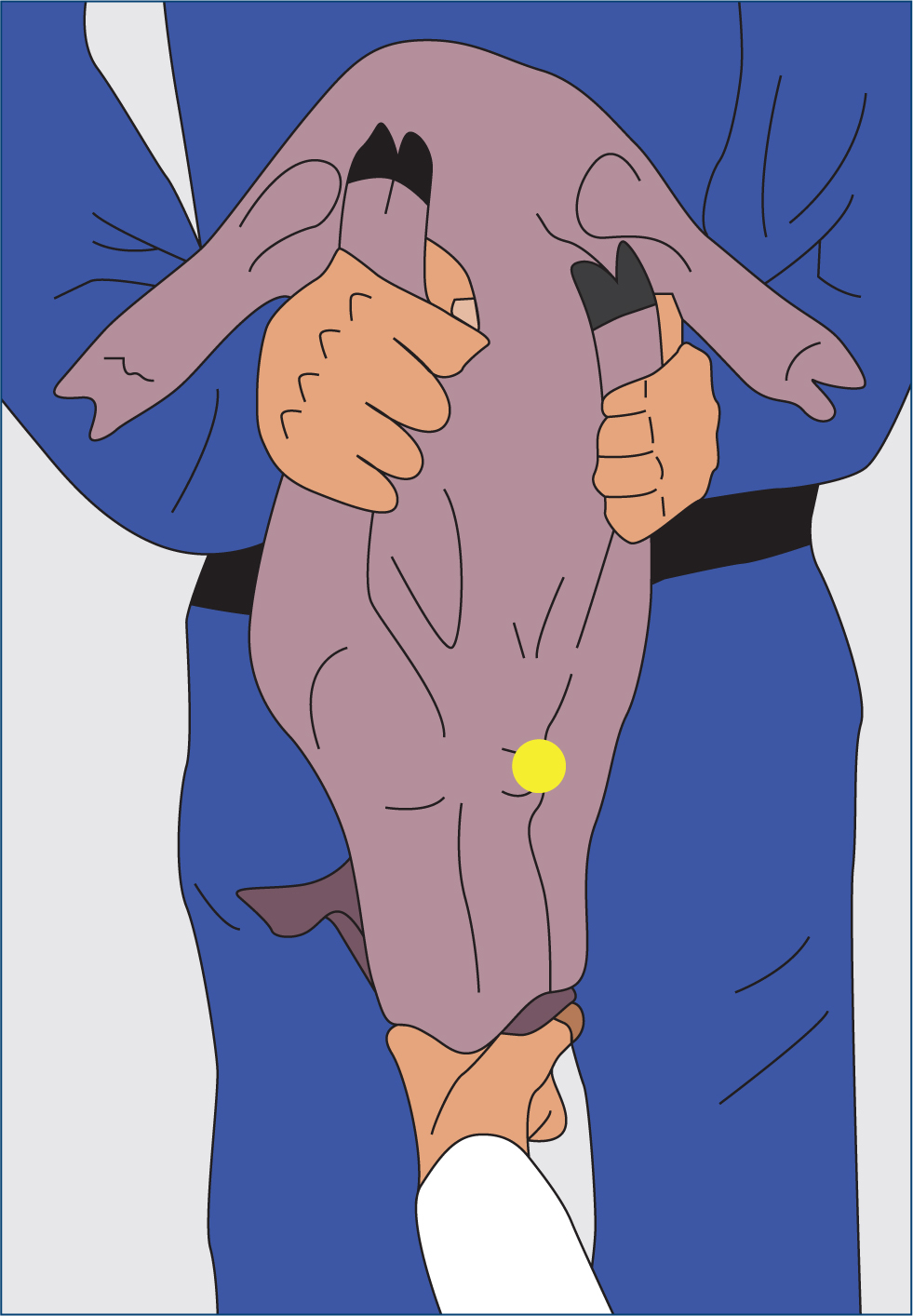
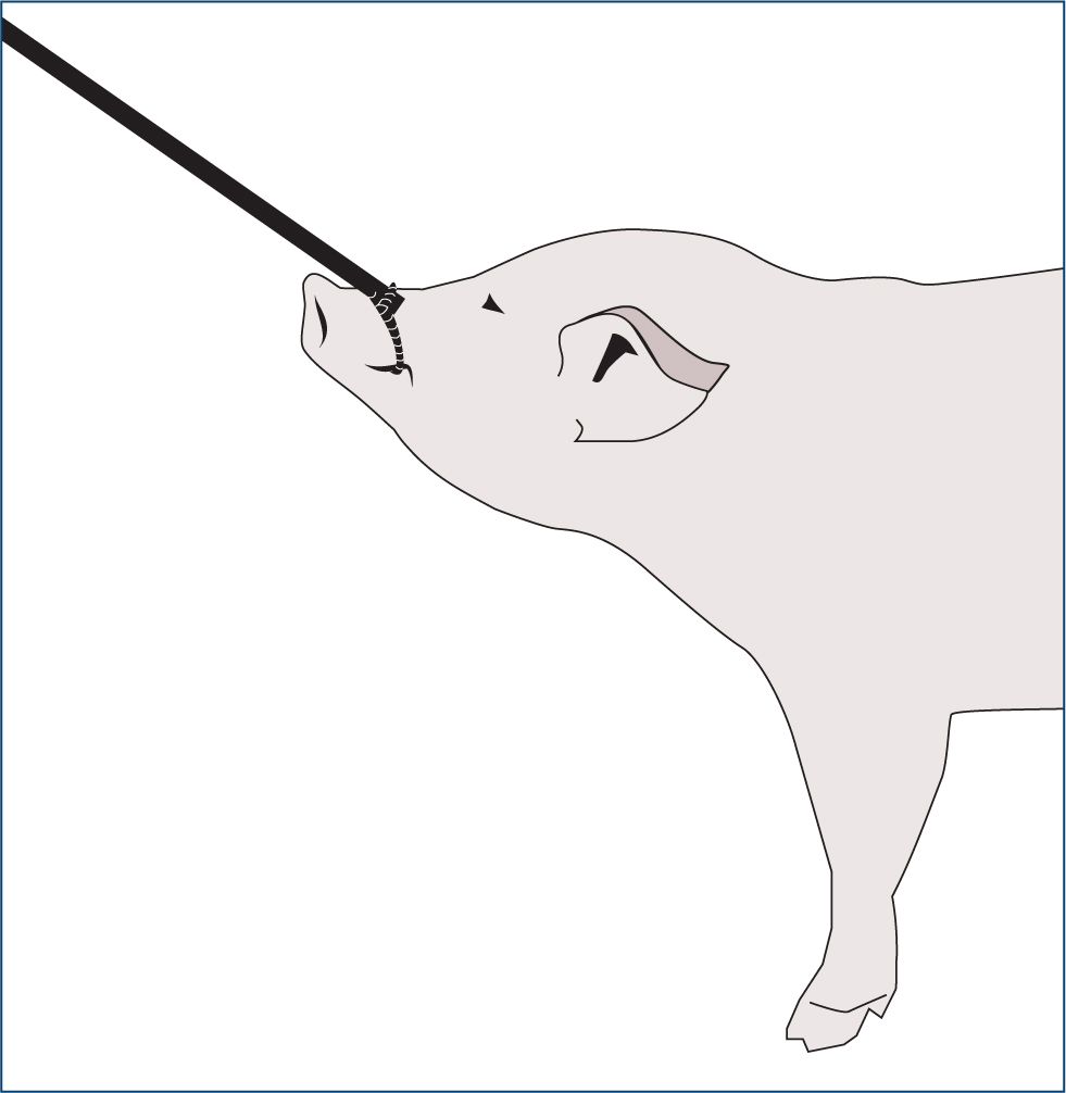
Pigs can be quite uncooperative when doing a clinical examination and this can be frustrating. They make a lot of noise and struggle, and as fast and strong animals they may push through people's legs and ram pig boards. They can also jump more than their height.
Male pigs especially can be extremely dangerous and if they are not intended for breeding, they should be castrated as a piglet under the supervision of a veterinary surgeon.
The auscultation area of the pig is small and this can be difficult, but the clinical examination should still involve taking the rectal temperature and handling the pig entirely.
If the pig is too uncooperative, it can be sedated with azaperone. Within the UK, there is a lack of effective sedatives and anaesthetics licenced for use in pigs. It would therefore be advisable to discuss options for effective sedation with a local pig veterinarian.
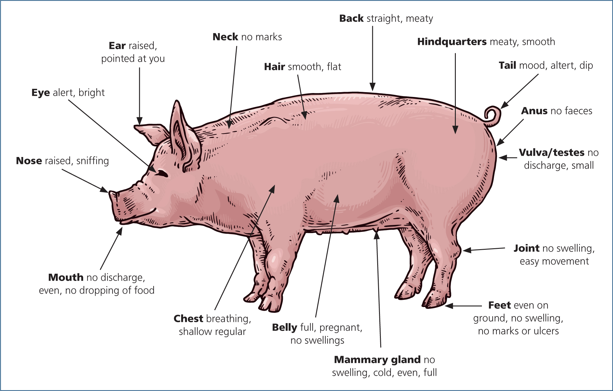
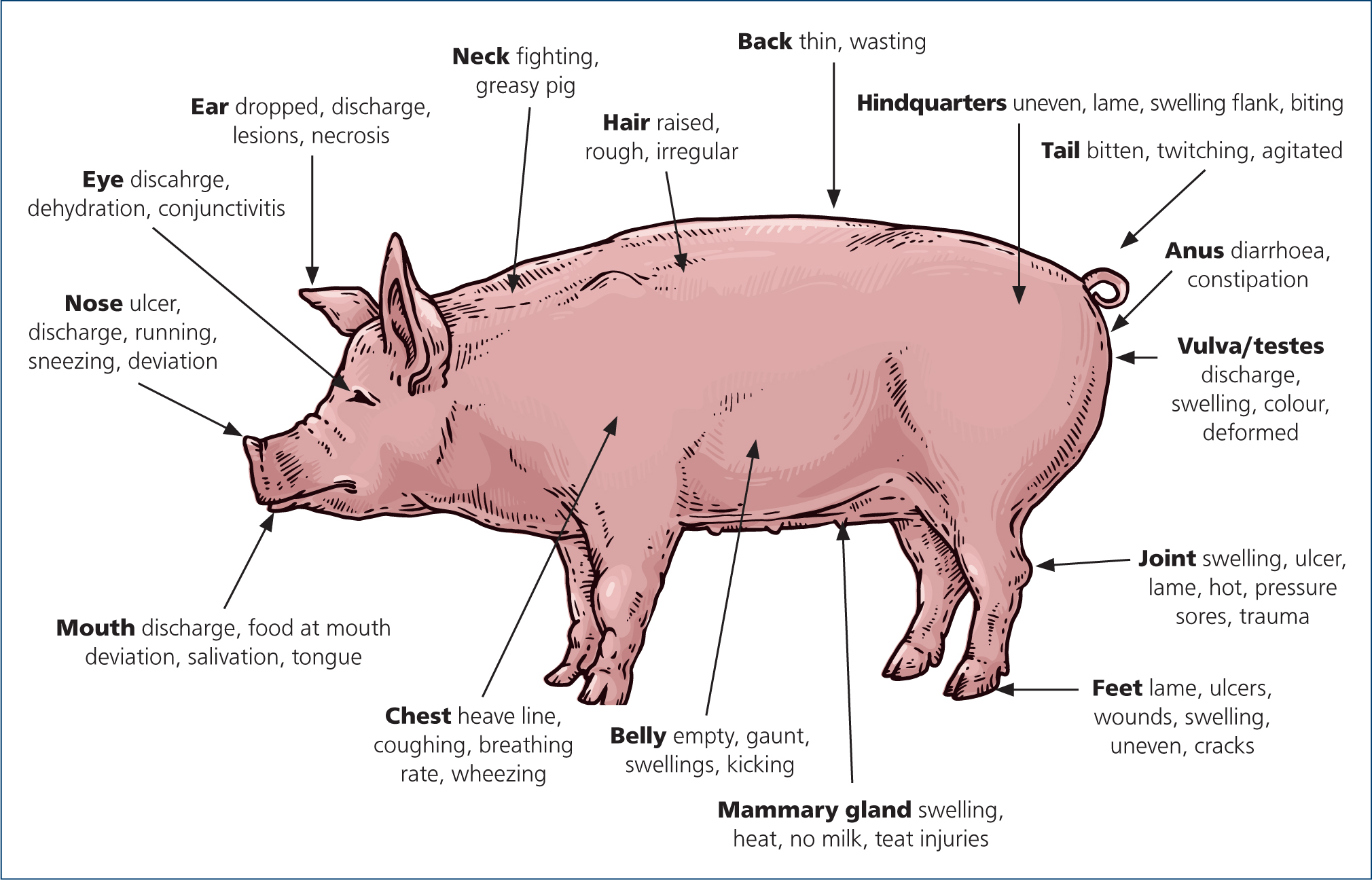
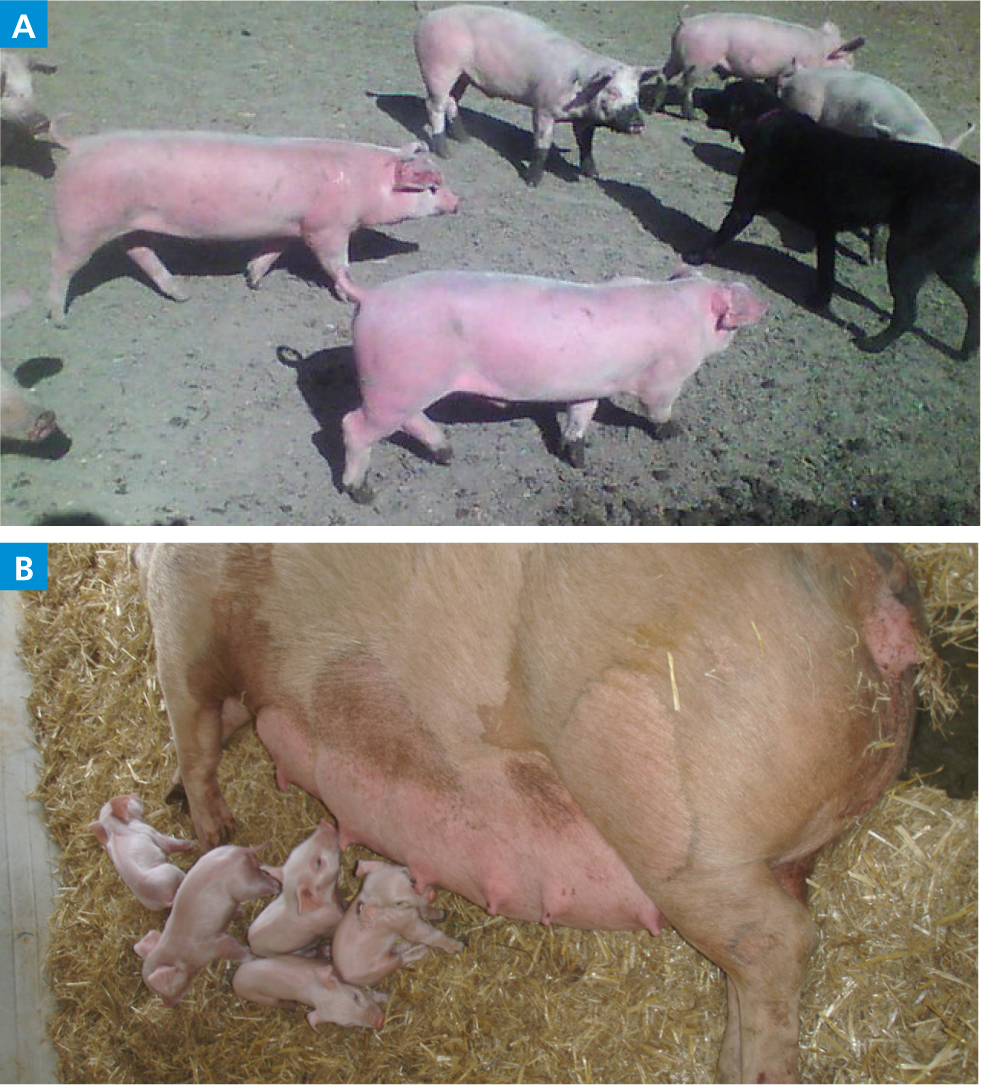
Refer to Figures 3 and 4 for the typical signs of healthy and unhealthy pigs (Swindle and Smith, 2016).
What breeds are common?
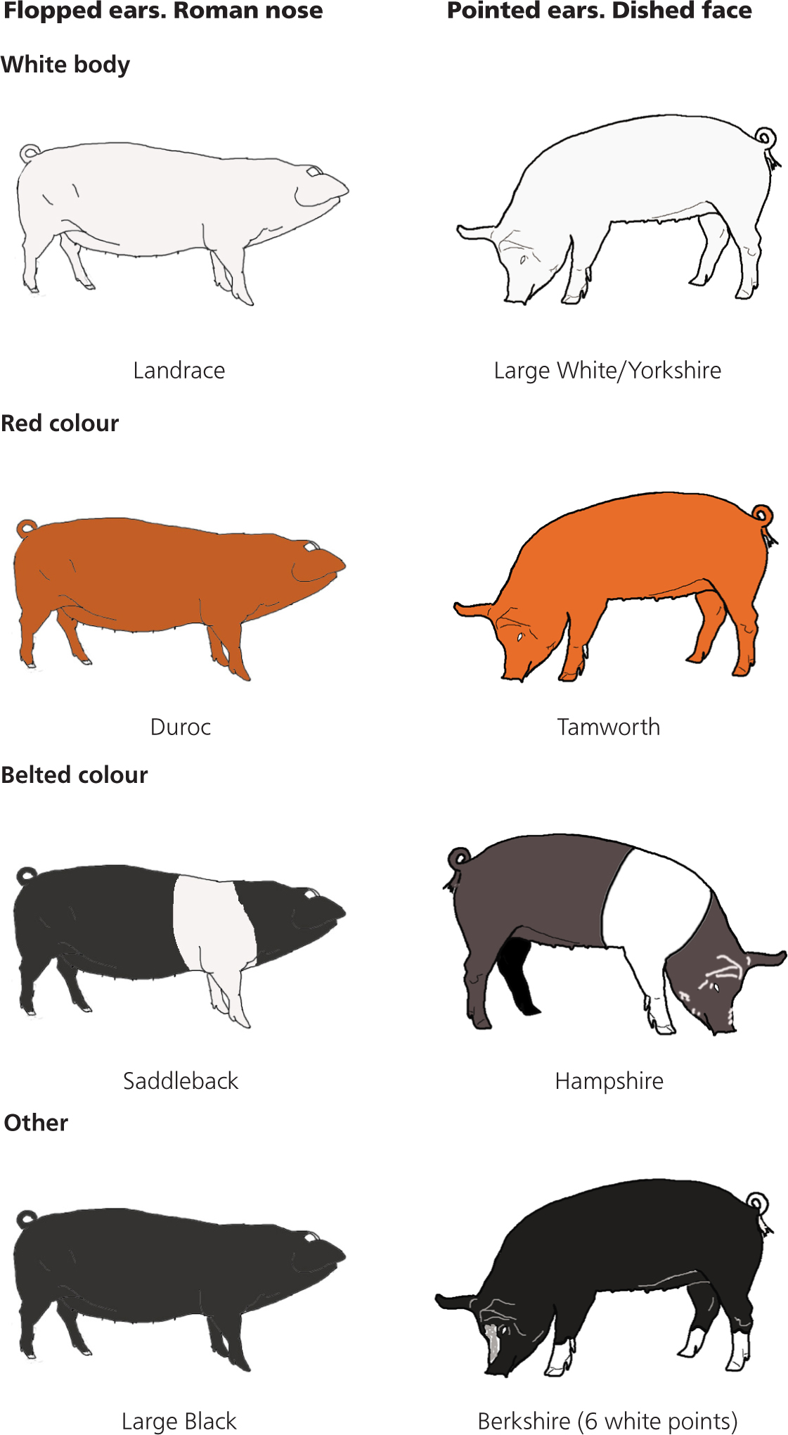
Within the UK there are a few breeds of pigs with which the veterinary surgeon should be familiar. UK pig breeds can be categorised into three by their colouring (white body, red colour and belted colour) and the position of their ears (Figure 5) (Shankland, 2011).
Common problems
Feed and water
While it may still be common practice, the feeding of kitchen waste to pigs is illegal. Vegetables can become cross-contaminated in the kitchen with meat products, so even ‘vegetable waste’ is not acceptable. Vegetables bought whole can be fed to pigs, but should not be fed as waste parts from kitchen preparation. As more food products are being imported from all over the world, there is an increasing likelihood that feeding cross-contaminated food waste can present highly infectious and aggressive pathogens to pigs and it is essential that we stop the introduction of African swine fever to UK pigs.
A guide to feed and water requirements for pigs:
- A growing pig will eat about 4% of its bodyweight per day of a balanced diet.
- A growing pig will drink about 10% of its body weight per day.
- For context, this means a growing 60kg pig will eat about 2.4 kg and drink 6 litres of water per day.
- Adult sows need to eat about 2.5kg per day and drink 12 litres.
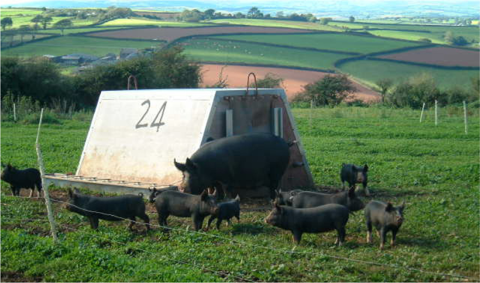
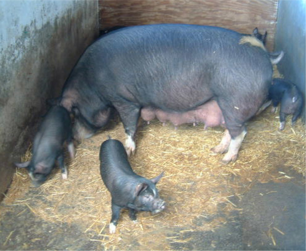
- A lactating sow on the other hand, will drink about 40–60 litres and eat up to 10 kg a day. (Figure 6) (Muirhead and Alexander, 2013; Carr et al, 2018; Department for Environment, Food and Rural Affairs, 2020).
Health issues
Abscessation
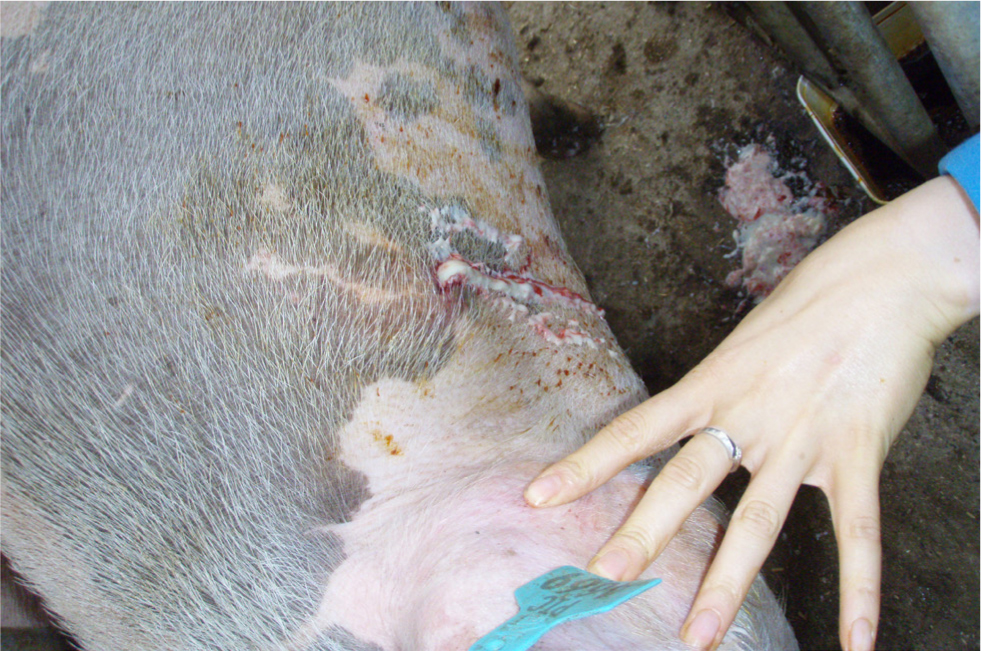
Pigs are prone to abscesses. Subcutaneous abscesses can be very large and may contain many litres of purulent material. After examination and confirmation of an abscess, lance the abscess to allow adequate drainage – no abscess pockets should be left (Figure 7). Flush with running water using a hose two to three times daily and provide pain management. Diluted hibiscrub with 5g salt per litre added can also be used to wash out the abscess. The causal organisms associated with abscessation are typically Streptococcus spp., Actinobacillus suis and Trueperella pyogenes. By the time the abscess is presented for treatment, the lesion may be sterile. Treatment using antibiotics such as lincomycin or a broad-spectrum antibiotic may be beneficial, but the abscess capsule will interfere with antibiotic dispersal.
Mange
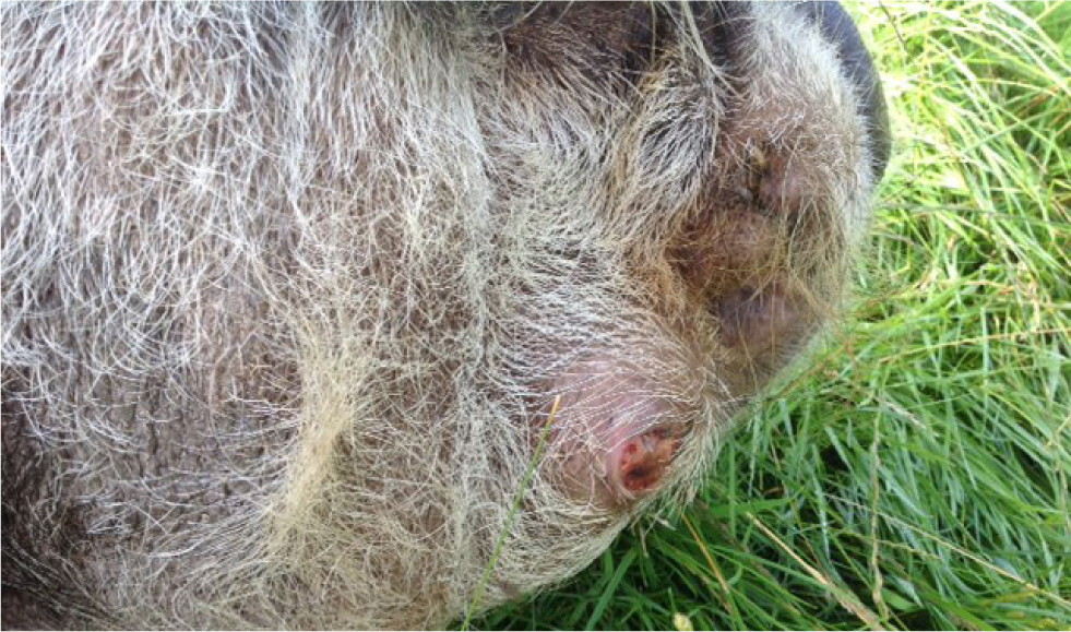
Sarcoptic mange is a serious parasitic infection of the pig caused by Sarcoptes scabiei var suis. The mite is able to survive for 21 days off the host in ideal situations. Mange will weaken the pig and is an added stressor. Affected pigs appear uncomfortable and will demonstrate intermittent body scratching, and the constant rubbing can also cause damage to buildings. Ear wax also increases, sometimes forming plaques. As the condition becomes chronic, hair will be rubbed off in places and the skin may become thickened (Figure 9). Mange affects pigs of all age groups, although sows and growing pigs most often exhibit the characteristic clinical signs.
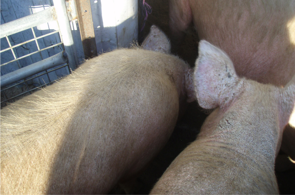
Avermectins can be adminstered for treatment via various routes, the most common being via injection or orally in feed. Mange should be considered a significant welfare problem and with the use of avermectins, it can be eliminated from all small-holdings.
Lice
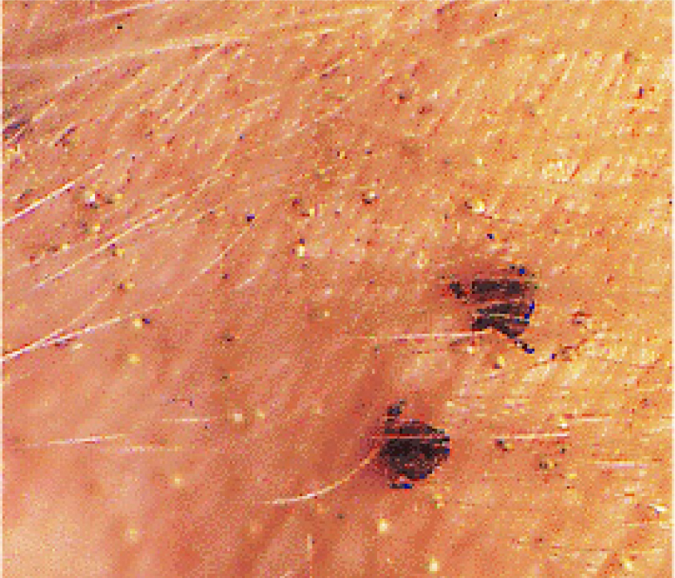
The pig biting louse is Haematopinus suis (Figure 10). These are the biggest lice known to man and thus they can be easily observed. Their life cycle occurs on the host's body and takes 30 days to complete, from egg to adult. However, the louse cannot live for more than 3 days away from the pig, theoretically making control easier than with mange, although in practice this has proven more difficult. It is possible that swine pox may be carried by lice. Lice are very sensitive to treatment with avermectins. To improve their efficacy, ensure that a two dose method is used with a 14 day gap between treatments.
Teeth and tusks
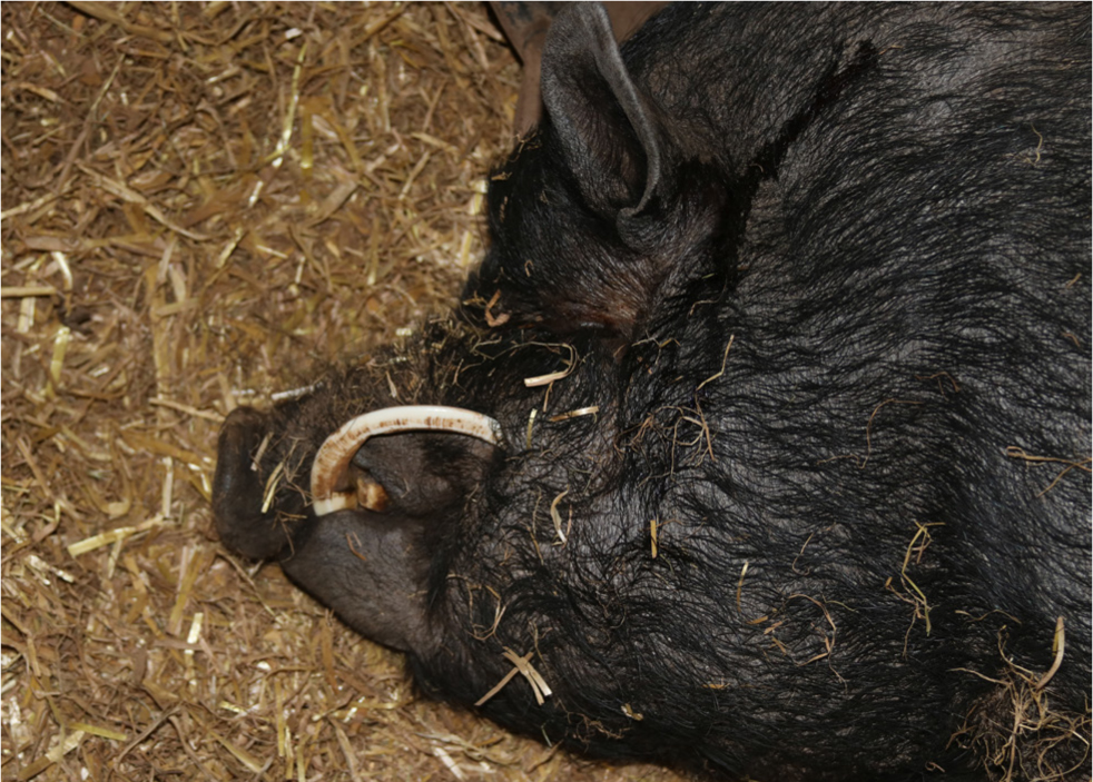
Pigs can develop a range of dental problems. Teeth impaction and abscessation can be a significant problem (Figure 8). Many sows have pain and difficulty eating, leading to reduced food intake. This may result in ill-thrift and even death. Tusks can become very large and pose a risk to people, other pigs and less commonly, may rotate back into the pig's face (Figure 11). Tusks can be trimmed using a Gigli wire or equine molar cutters and under suitable sedation. Pigs can also be provided with a large rock or brick so they can keep their tusks trimmed (Duncanson, 2013).
Erysipelas
Diamond lesions of erysipelas can be felt and seen on the back of the pig (Figure 12).
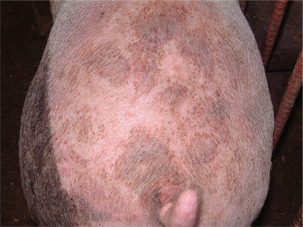
Erysipelas of pigs is caused by the bacterium Erysipelothrix rhusiopathiae, which is commonly found in soil samples. It can therefore enter the small holding through the feed, water supply or general rooting. When grain is harvested, 1–2% of the weight is soil. The bacterium can also be found in fish, and fishmeal can be a source of infection. Erysipelothrix rhusiopathiae can also cause problems in turkeys and sheep, which can then cross-infect pigs on multispecies farms. In addition to this, up to 20% of the pigs on a farm may carry the organism on their tonsils without presenting with any clinical signs.
The organism can gain access to the pig via many routes. Classically, most infections are oral and come from contaminated feed and/or water. In acute cases, the pathogen enters the blood stream via the pharynx. The diamond lesions are actually the consequence of microthrombi and bacterial emboli in dermal capillaries and venules, which may lead to circulatory stasis and necrosis. Usually, affected pigs have already been sick for 2–3 days before the lesions become large enough to be visible. In the chronic form, erysipelas arthritis can develop as a result of swelling and inflammation damaging the joint surfaces. This can take months to develop, so securing a definitive diagnosis can be difficult as the lesions are sterile by the time the clinical signs are recognised.
While diamond lesions are not definitive, they are very suggestive as the major visible and palpable clinical sign of erysipelas infection. Suspected pigs should be treated with penicillin immediately. If there is a significant improvement within 24 hours, this is very suggestive of erysipelas. Vaccination is advised for all adult pigs every 6 months.
It is worth noting that Erysipelothrix rhusiopathiae is a zoonotic pathogen and it is therefore essential that all those involved in treating and caring for affected pigs should wash their hands regularly.
Swine influenza
Swine influenza virus (SIV) is an orthoinfluenza type A virus. This is an enveloped ribonucleic acid (RNA) virus. It has eight segments to its genome and this allows for reassortment and recombination of the genome. Three major subtypes of SIV infecting swine have been described (H1N1, H1N2 and H3N2), each causing similar clinical signs and lesions. Other types can be found worldwide.
Typical SIV infection is described as acute outbreaks characterised by sneezing and nasal discharges, a non-productive cough, tachypnoea, fever, anorexia and a reluctance to move. The clinical signs spread around the small holding rapidly and occur in multiple age groups. SIV infections can also cause reproductive problems, as the high temperatures experienced can cause pregnant sows to abort their fetuses.
The recommended treatment on a small holder is to provide supportive therapy. Antimicrobials may be considered to control secondary infections. Antipyretics will assist recovery and the resulting pyrexia.
It is important to remember that swine influenza may pass from humans to pigs and vice versa, hence precautionary measures should be taken.
Piglet diarrhoea
Escherichia coli (E. coli) is an extremely important bacteria when considering the health of pigs. The majority of serotypes are beneficial and even vital for the normal function of the microflora of the pig and its metagenomics. However, a few strains are also associated with a variety of conditions from toxaemia in very young pigs, to diarrhoea in pre- and post-weaning pigs. E. coli are not significant pathogens in pigs older than 10 weeks of age. The pathogenic E. coli tend to be haemolytic (as they are short of iron) and this can be used as part of an initial screening for the cause of diarrhoea.
Most E. coli problems are associated with management and environmental factors, in particular chilling draughts and sanitation. Examining the environment is an essential component to any diagnosis. E. coli-induced diarrhoea is more prevalent in parity 1 litters and can play a role in causing diarrhoea in the pig in three distinct phases.
- 0–3 days of age: piglets present with watery yellow diarrhoea and often sudden death.
- 4–10 days of age: piglets present with a pasty yellow coloured faeces. In the early stages of the clinical signs, some vomiting may also be recognised. Some piglets may be found dead, but most have clinical signs that lead to dehydration and ultimately death. If diarrhoea occurs after day 10, but before weaning, coccidiosis should be considered as part of the differential diagnosis.
- Post-weaning (normally starts after 3–5 days): pigs present with both acute and chronic diarrhoea. The diarrhoea progressively leads to dehydration and death. Weaners may demonstrate ill thrift.
It is important to treat the whole litter as soon as one piglet starts to show clinical signs. Place a trough drinker filled with water, electrolytes and glucose. In emergencies, the use of cola drinks or lemonade may also be very beneficial. This must be replaced at least four times daily.
Stockpersons should wash their hands after treating the piglets and dip their boots in disinfectant or, ideally, wear gloves and have different boots for each affected room/batch. Each brush and shovel should be placed in disinfectant when not in use. Ensure disinfectant is still active. Ensure that the tools and environment are thoroughly cleaned between batches. Consider lime washing to enhance hygiene and disinfection.
Following an antibiogram, suitable antibiotic therapy may be required. Note that salmonellosis may also look like severe E. coli and should always be considered in the differential list.
Ascaris worms
The adult Ascaris suum worm is large, at about 20–40 cm long (Figure 15). An adult female lives about 6 months and may lay 2 million eggs per day, but egg production is extremely variable day-to-day. The eggs are very infectious as they are extremely sticky and will be easily transported onto the farm by pigs, insects, birds and equipment. They are extremely resistant and can survive for more than 7 years in the environment. Generally, disinfectants have little effect on the eggs. There are no obvious clinical signs but the adult worms can be seen in the faecal stool.
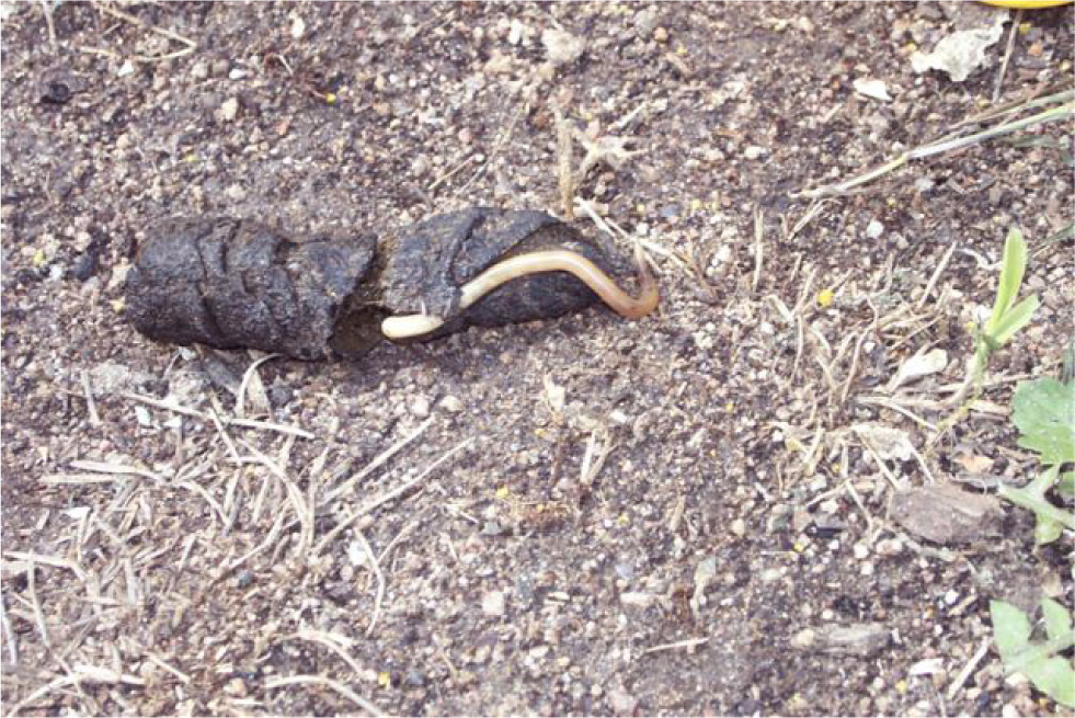
The migrating larvae passing through the liver may result in temporary scarring. This might be seen in the slaughterhouse as white spot liver and lead to condemnation of the liver. As the larvae pass through the lung, there may be a soft cough.
Sudden death: torsion and poisoning
A common cause of sudden death in pigs is an abdominal catastrophe, characterised by a torsion or twist of the stomach or intestines (Figure 16). The twist is commonly associated with the intestinal tract at the mesentery root or is associated with a mesenteric tear. Occasionally, the spleen or a liver lobe can become associated with a twist. Diagnosis is made at post-mortem. With scrotal or umbilical hernias, intestinal entrapment is common.
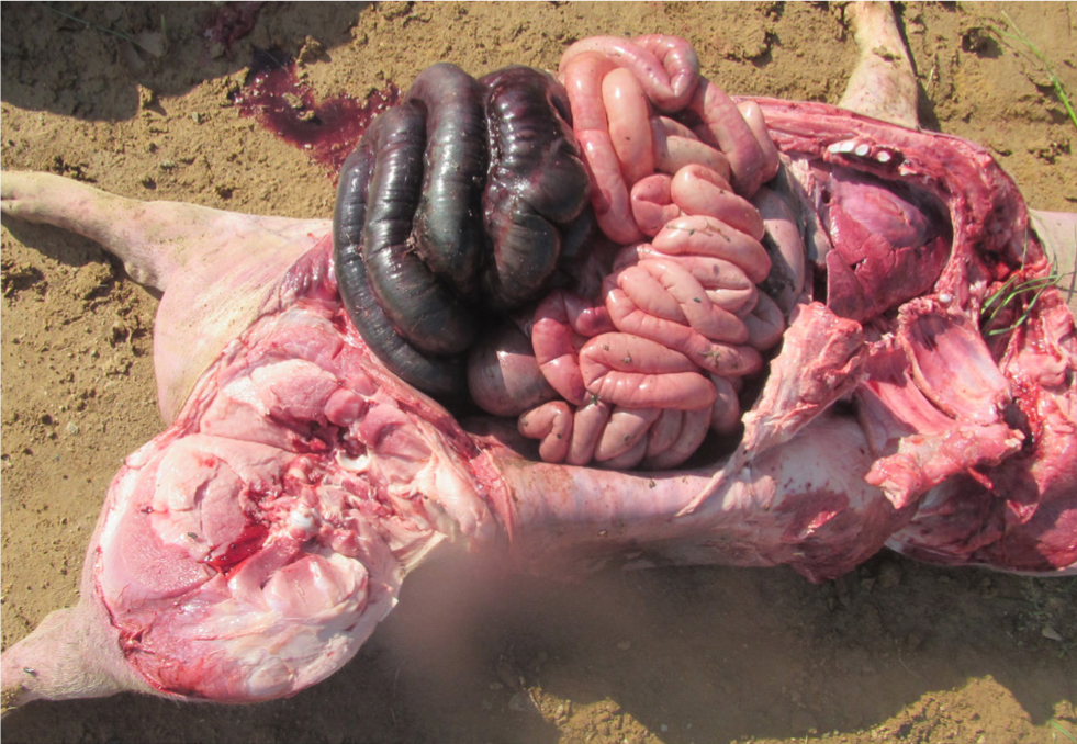
Torsions may be associated with a change in feeding routines or feeding interruptions. Therefore, variation in the time of feeding should be avoided, particularly over the weekend, in the adult herd. Ensure feeder storage capacity is sufficient for overnight, in case of feed delivery interruption.
Intestinal perforation is uncommon, but can result in sudden death associated with release of abdominal contents into the peritoneum causing peritonitis. The ingestion of hard plastic brush bristles have been found and determined as the cause of this in a few cases.
Constipation
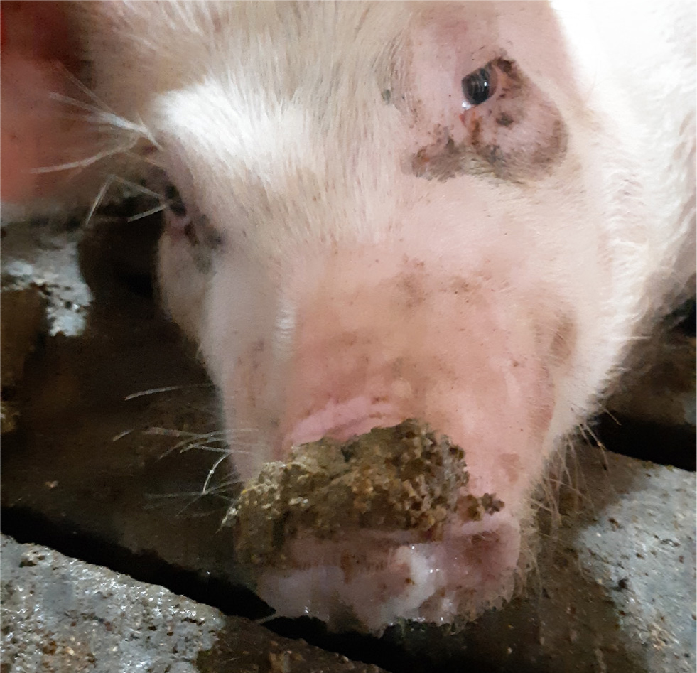
Constipation in sows is a particular problem in the periparturient phase. It results in difficulties with parturition and subsequent milk production, especially of colostrum. The sow presents with small hard round pellets of faeces (Figure 17). These can become impacted in the rectum, reducing the available space in the pelvis and thus increasing the risk of stillborn piglets. The absorbed endotoxin reduces prolactin production, reducing milk output, which can appear as agalactia. It is generally advised to feed extra fibre or bran for a day or two, pre-farrowing, to assist with defecation.
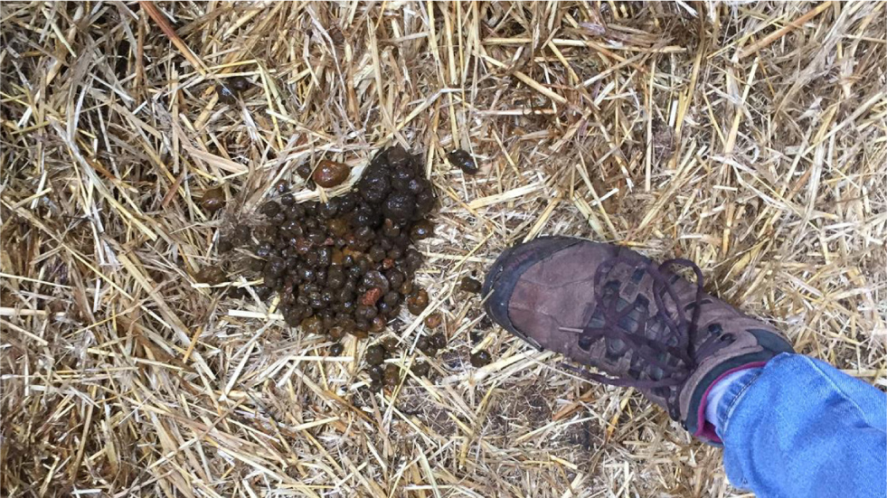
Table 1. Zoonotic diseases of pigs
| Anthrax |
| Brucellosis |
| Campylobacter jejuni |
| Chagas' Disease – Trypanosoma cruzi |
| Chlamydia |
| Clostridium perfringens type A |
| Ebola Reston |
| Erysipelas |
| Escherichia coli |
| Hepatitis E virus |
| Japanese B encephalitis |
| Louping ill |
| Leptospirosis |
| Meliosis – Burkholderia pseudomallei |
| Nipah disease |
| Pasteurellosis |
| Rabies |
| Ringworm |
| Salmonellosis |
| Streptococcus suis II |
| Spirometra erinacei |
| Swine Influenza |
| Taenia solium |
| Toxoplasmosis |
| Trichinella spiralis |
| Tuberculosis |
| Vesicular diseases |
| Yersina enterocolitica |
Constipation can be extremely severe in pigs when it results in impaction of the colon, which may also become life-threatening. Medication with enemas and oral laxatives, such as mineral oil or magnesium sulphate (5 g/100 kg) to soften the faeces, is recommended. If the pig is very dehydrated, several litres of fluid may be administered through the rectum. Constipation is a serious condition and needs to be treated as an emergency.
Parvovirus
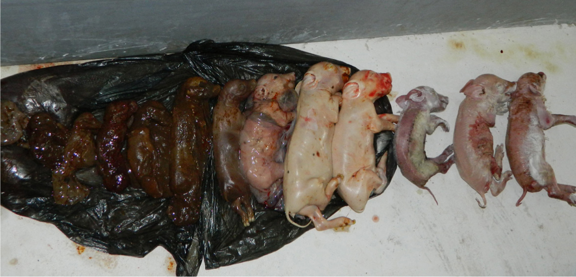
Porcine parvovirus causes reproductive problems, the most obvious of which being an acutely increased number of mummified piglets, particularly in non-vaccinated gilts and sows (Figure 18). Embryonic or foetal death results from transplacental infection with parvovirus. The source of the virus is faeces and other body secretions from an infected animal contaminating the environment. Virus can also be introduced via fomites or rodents. The virus is very hardy and can survive for months in the environment.
Vaccination of gilts is the control method of choice for porcine parvovirus. Maternal antibodies can interfere with the immune response of gilts to vaccination and these antibodies may be present until 6 months of age. Therefore, incoming gilts need to be vaccinated after this age and at least 2 weeks before breeding. There is no requirement to vaccinate boars.
Heat-related environmental problems
Sunburnt pigs can be severely affected. They can become ataxic and stiff, possibly becoming prostrate, tachypnoeic and eventually dying. In males, this may affect the testes and often the dorsal aspect, resulting in infertility.
Providing adequate, well-ventilated shade and wallows to outdoor pigs and monitoring the impact of the sun in indoor housed pigs is paramount to maintaining their health. In pigs affected by sun exposure, keep them cool with water, as pigs cannot sweat to cool themselves. In small numbers, the pig skin can be protected with sunscreen and ‘after-sun’ oils.
Big issues
African swine fever:
African swine fever virus is the only representative of the Asfarviridae family. It is one of the largest and most complex viruses and is able to cause fatal disease in pigs of all breeds and ages. The current pandemic of African swine fever is associated with type II.
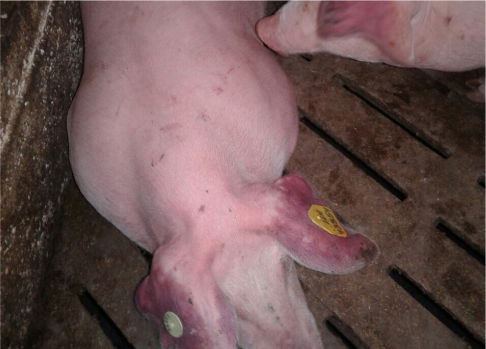
Although wild African suids are resistant to the pathogen, domestic pigs and European wild boar can develop a number of clinical signs. Mortality is the usual outcome for those infected with highly virulent strains. Acutely affected animals develop a high fever, often becoming very quiet and reluctant to move. Many pigs may vomit and present with dyspnoea (as a result of severe pulmonary oedema). Pregnant sows will abort and lactating sows will stop lactating. In most cases, a haemorrhagic diathesis develops, with potential cutaneous haemorrhages, epistaxis and bloody diarrhoea. If the pig has red ears and is very pyrexic (Figure 20), please contact the government veterinary services (Department for Environment, Food and Rural Affairs, 2020).
Foot and mouth disease
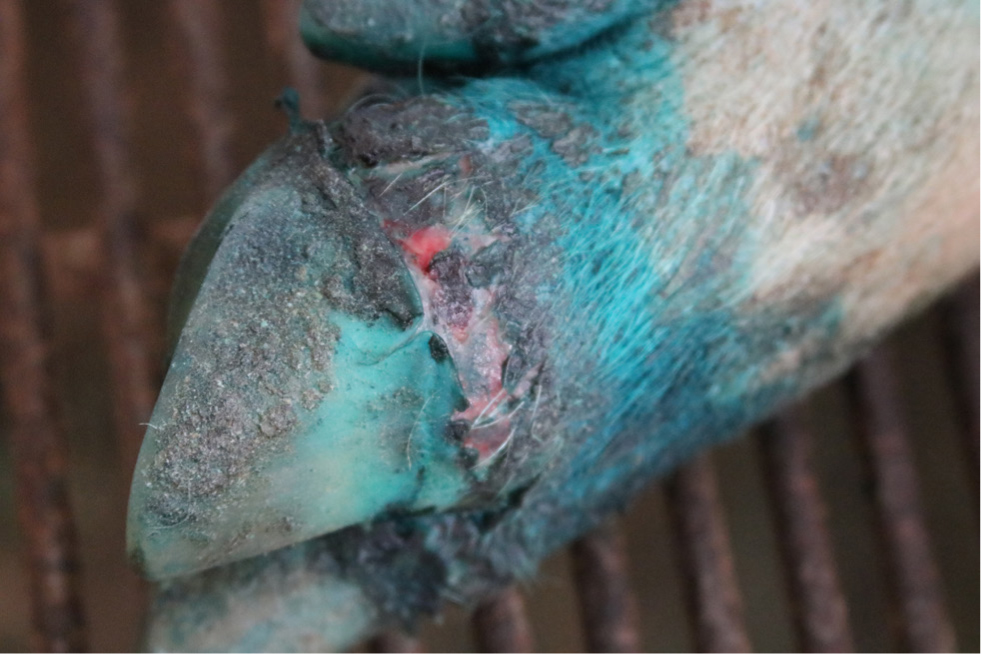
Foot and mouth disease is caused by an Aphthovirus of the Picornoviridae family. Pigs of all ages can be affected in the initial outbreak. In an outbreak, the clinical signs may not be seen for 1–5 days, and can take as long as 21 days to develop. The major clinical sign is that several pigs are suddenly lame. When the animals are clinically examined, the skin around the snout lips, tongue, inside the mouth, around the coronary band and the soft skin on the feet becomes whiter (blanched). Then, within hours, vesicles (blisters) may develop. These may also be seen on the sows' teats. The vesicles are rarely seen as they rupture within 24 hours and secondary infection of the exposed skin occurs. Over a few days, in some pigs the hoof may become detached, revealing the painful raw tissues underneath (Figure 21). The pigs may have pyrexia of 40.5°C. If there is any suspicion, contact the government veterinary services (Department for Environment, Food and Rural Affairs, 2020).
Needles and medication
Intramuscular injection is the easiest method of administering an injection. Subcutaneous injection in the pig is difficult as they have subcutaneous fat and the skin is closely applied to underlying tissues. Ensure the syringe and needles are strong. Appropriate needle sizes are outlined in Table 2. There is increasing use of needless injection technologies, at present this is mainly for vaccination (Carr and Smith, 2013).
Table 2. Appropriate needle sizes for pigs
| Weight of the pig | Diameter mm | Length mm |
|---|---|---|
| 1–7 kg | 0.8 | 16 |
| 7–60 kg | 1.1 | 25 |
| 60–110 kg | 1.3 | 25 |
| Adults | 1.3 | 40 |
Site of injection
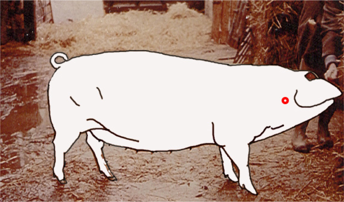
An intramuscular injection can be administered just behind the ear (Figure 22). Rub the pig's ear and insert the needle and inject quickly. To administer medication by injection it be best to have the pig restrained. Note that other sites are unsuitable, especially if the pig is intended for human consumption.
However, if the pig is eating then medication can easily be administered orally in water or using tablets. If the medication is disguised in food, such as grapes, apple or a little chocolate, the pig will often digest the medication with gusto!
Vaccination and preventative medicine
Avermectins can be used to treat internal and external parasites. It is important that backyard pigs should remain free from infections with Sarcoptes scabiei and Haematopinus suis, as both lice and mange are considered a welfare problem in pigs. This can be achieved by an initial eradication programme, with any new animals being isolated and medicated before being introduced into the farm. This elimination method removes the need for regular preventative medication. Internal parasites can be medicated as the need arises and routine medication should be avoided, as internal parasites are normal in free-range pigs. Unless the parasite becomes a problem, the pig and parasite should be allowed to reach a natural equilibrium, although their general health should always be maintained. The unnecessary use of antimicrobials only increases the risk of resistance developing. Adult pigs should be routinely vaccinated against erysipelas twice a year.
Before their first pregnancy, gilts should be vaccinated against parvovirus. Before farrowing, it may be worth considering an E.coli vaccination at 6 and 3 weeks pre-farrowing. Other pathogens can be vaccinated against as the size of a small holding or sanctuary increases, but if the mother population is made up of less than ten sows then concentrating on erysipelas should be sufficient unless any clinical signs observed dictate the presence of other pathogens.
KEY POINTS
- Communication between vets and owners is important, it can be helpful to request a short video (around 30 seconds) of the pig(s) before attending the farm.
- Pigs can be very stubborn and make a lot of noise.
- The feeding of kitchen waste is illegal and a visit is always a great opportunity to educate owners.
- Most clinical presentations in pigs are basic issues and can be resolved easily, but if you have concerns ask your colleagues from the Pig Veterinary Society.

