Rehabilitating the canine patient from spinal cord injury is somewhat more complicated than with patients affected by joint disease, as there is often neurological impairment, as well as pain and weakness, to address. Before referral, most patients will have had a detailed neurological examination carried out by the veterinary surgeon, with use of diagnostic imaging modalities to enable diagnosis, followed by treatment which may or may not include surgery.
With any rehabilitation programme, the patient must first be assessed to gather information and allow the therapist to create a problem list. This includes identifying the location and degree of any neurological impairment, assessing whether the patient requires additional pain management and identifying any areas of muscular weakness that could be strengthened by use of physical therapy techniques.
Unlike the other articles in this series, where a more generalised approach to the stages of rehabilitation could be applied, this article discusses the specific rehabilitation therapies which may assist with recovery in the ambulatory and non-ambulatory patient, including physical therapies, electrotherapies and acupuncture. Neurological conditions affecting the cervical vertebrae, resulting in tetraparesis, are often managed with hospitalisation and intensive therapy and are thus outside the scope of this article.
Addressing neurological deficits
It has been demonstrated that the use of rehabilitation therapy following spinal cord injury improves a patient's return to function, particularly if it is initiated in the first 2 weeks (Sumida et al, 2001). Physical therapy not only stimulates strengthening and regeneration of muscle tissue following neurogenic atrophy, it also reinforces corticospinal pathway connections to re-educate motor and sensory abilities (Frank and Roynard, 2018).
The increased use of rehabilitation therapies could also reduce recovery times, with a recent study demonstrating a decrease in time to ambulation following surgery for thoracolumbar disc extrusion, from around 25 to 14 days in the patient group which received physiotherapy and photobiomodulation, compared with those receiving physiotherapy alone (Bruno et al, 2020).
In the initial assessment by the physiotherapist following referral, a detailed neurological exam is conducted, during which the therapist will assess the following:
- The presence of deep pain
- Reflexes
- Evaluation of posture
- Gait analysis
- Proprioceptive awareness.
Patients can then be grossly divided into two categories: ambulatory and non-ambulatory.
The focus of the acute phase following injury for non-ambulatory patients is on maintaining musculoskeletal health. This is done by re-educating patients on how to perform basic functional tasks (Drum, 2010) such as standing, performing transitions from lie-to-sit-to-stand and early ambulation. This retraining is task specific, so emphasis should be placed on ensuring the patient can perform each individual transition and activity required for everyday function. Proprioceptive enrichment is also particularly important to these patients as it is key to neurological recovery. Examples of exercises which would be included in a rehabilitation session at this stage are detailed in Table 1.
Table 1. Exercises to aid the canine patient's rehabilitation from a spinal injury
| Exercise | Description | Benefit to patient |
|---|---|---|
| Passive range of motion | The therapist moves the joints of the patient's limbs individually from digits to hip/shoulder slowly and smoothly, through flexion and extension, until natural resistance is felt. This is done around 10 times, 3–4 times daily |
|
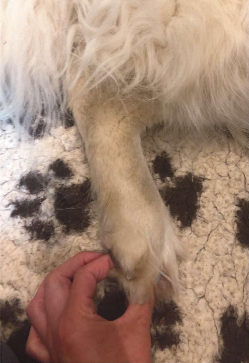
| Active range of motion | The toes of the patient are stimulated (Figure 1) to trigger a flexor withdrawal response. This is done 3–5 times on each foot, 3–4 times daily |
|
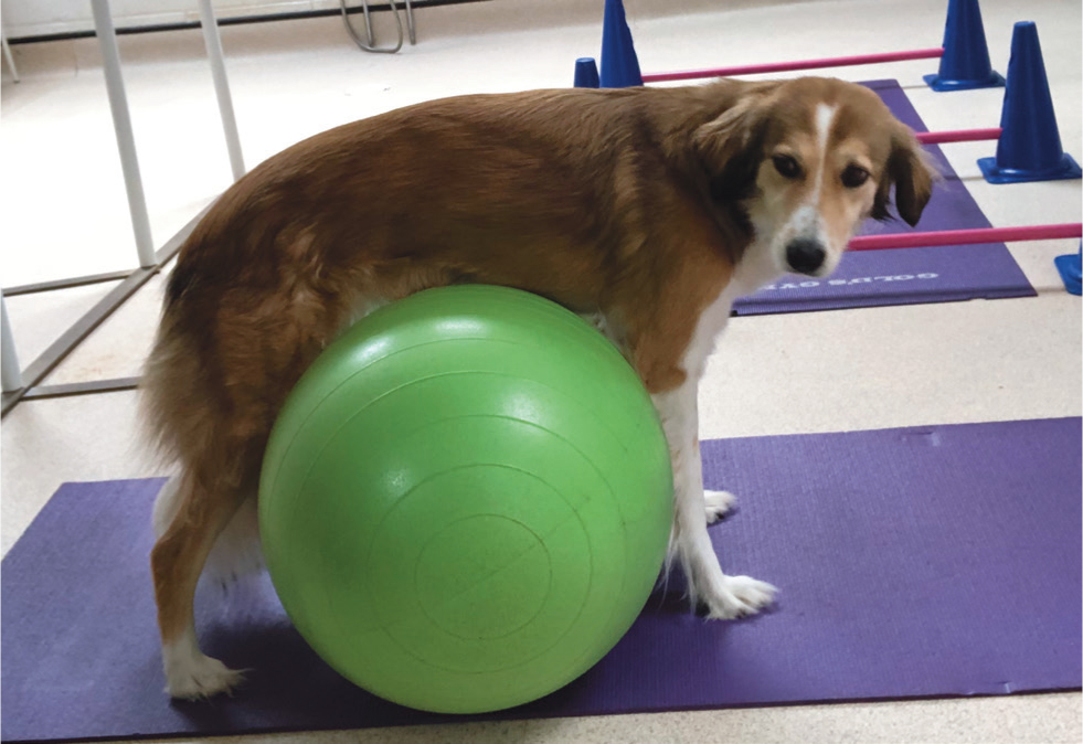
| Facilitated standing | The therapist assists the patient into a standing position. Depending on patient size, this is done using either support from the therapist alone, with the assistance of aids such as a sling, peanut (Figure 2) or physio ball (it is important to check foot position to ensure it is normal with no knuckling). The patient is encouraged to stay in this position, initially for 15 seconds, building gradually, 3–4 times daily |
|
| Rhythmical stabilisations | While the patient is standing, gentle pulses are applied to shift the patient's weight on and off the hind quarters, starting with 15 seconds and building gradually, 3–4 times daily |
|
| Sit-to-stand exercises | The patient is assisted into a sit position (supported onto the therapist's knee) and then raised into a standing position, initially for three repetitions, then increased gradually, 3–4 times daily |
|
| Aquatic treadmill | The therapist uses gait patterning techniques to encourage early ambulation of the patient on the treadmill belt, while the buoyancy of the water assists in supporting the patient's bodyweight. Initial sessions may involve 3–4 sets of 1 minute, building gradually. Aquatic therapy may be used daily if available while the patient is hospitalised |
|
For the non-ambulatory patient, nursing care is of prime importance during the early postoperative period. Prevention of pressure ulcers through regular turning of the patient and provision of deep, supportive bedding is necessary, as well as the prevention of urine scald through use of regular catheterization, bladder expression and removal of soiled bedding. These factors can have a huge impact on patient welfare (Elphee, 2011).
Patients who are ambulatory but weakened, and have ongoing neurological deficits, will benefit from the physical therapy techniques outlined below, with additional exercises to strengthen the limbs and enhance proprioception further:
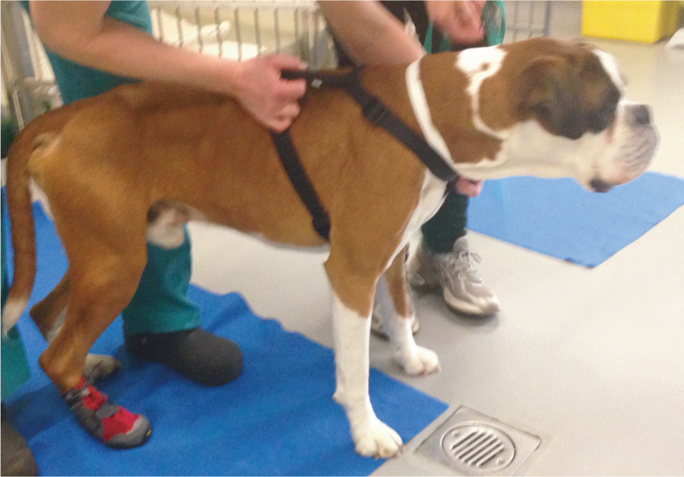
- Short periods of facilitated gait on land, with the use of a sling or harness for support (Figure 3). This should be performed on a non-slip surface 3–5 times daily. The use of non-slip, lightweight boots (Figure 4) may be essential to prevent the development of sores on the feet during knuckling/scuffing. Walking should be carried out at a slow pace and may be facilitated with the use of a harness or lead, to give the therapist control over speed. This allows for careful consideration to be given to foot placement and for assistance to be provided, where the patient is unable to place correctly (Drum, 2010).
- When the patient becomes more advanced and does not require assistance during gait, the addition of low cavaletti poles to encourage the patient to lift and place their feet is beneficial. As the recovery progresses, walking on uneven surfaces and gentle uphill slopes are excellent ways to enhance proprioception and strengthen the limbs and core.
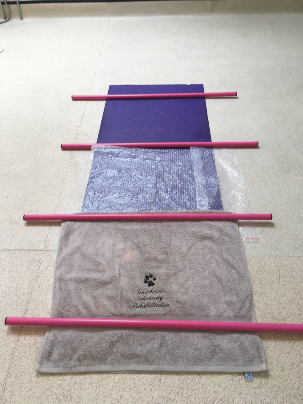
- Setting up a proprioceptive track (Figure 5) using at least three different non-slip surfaces (examples include carpet, rubber mat, sand, grass and bubble wrap) and walking the patient over it in a slow, controlled fashion, is an excellent way of enhancing proprioception and helping the patient improve their coordination.
- Continuing hydrotherapy, with increasing speed and duration, assists with muscle strengthening, regaining cardiovascular fitness and proprioception.

Electrotherapy
Neuro-muscular electrical stimulation involves placing electrode pads over a specific muscle, through which a current is applied, evoking enhanced force contraction, which is the ability of the muscle to contract (Canapp, 2007). It is widely used in both human and animal neurological patients, to minimise disuse atrophy, stimulate improvements in the patient's gait and improve motor control (Frank and Roynard, 2018). Studies have shown the application of neuro-muscular electrical stimulation can be hugely beneficial to the neurological patient, when used together with physical therapies rather than alone (Harvey, 2016). This reiterates the commonly accepted theory that a multimodal approach to rehabilitation is the most beneficial to the patient. It should be noted that neuro-muscular electrical stimulation should be used with caution in patients displaying increased (spastic) muscle tone (Millis and Levine, 2014).
The magnetic field produced as a result of pulsed magnetic field therapy (Figure 6) has a variety of effects on the tissues via the activation of biological processes. This includes increases in cell membrane potential, tissue oxygenation, vasodilation and analgesia (Pinna et al, 2013), making it an appropriate therapy to assist with healing and pain management in dogs suffering from spinal cord injury. In a study of patients post-hemilaminectomy surgery, it was revealed that dogs receiving pulsed magnetic field therapy had improved wound scores at 6 weeks postoperation, as well as improved owner perceptions of pain, subsequently leading to a less frequent administration of analgesia (Alvarez et al, 2019). Another study confirmed these effects on lower incision-related pain scores and improved proprioception (Zidan et al, 2018), but further studies are warranted to fully evaluate the effects of pulsed magnetic field therapy.
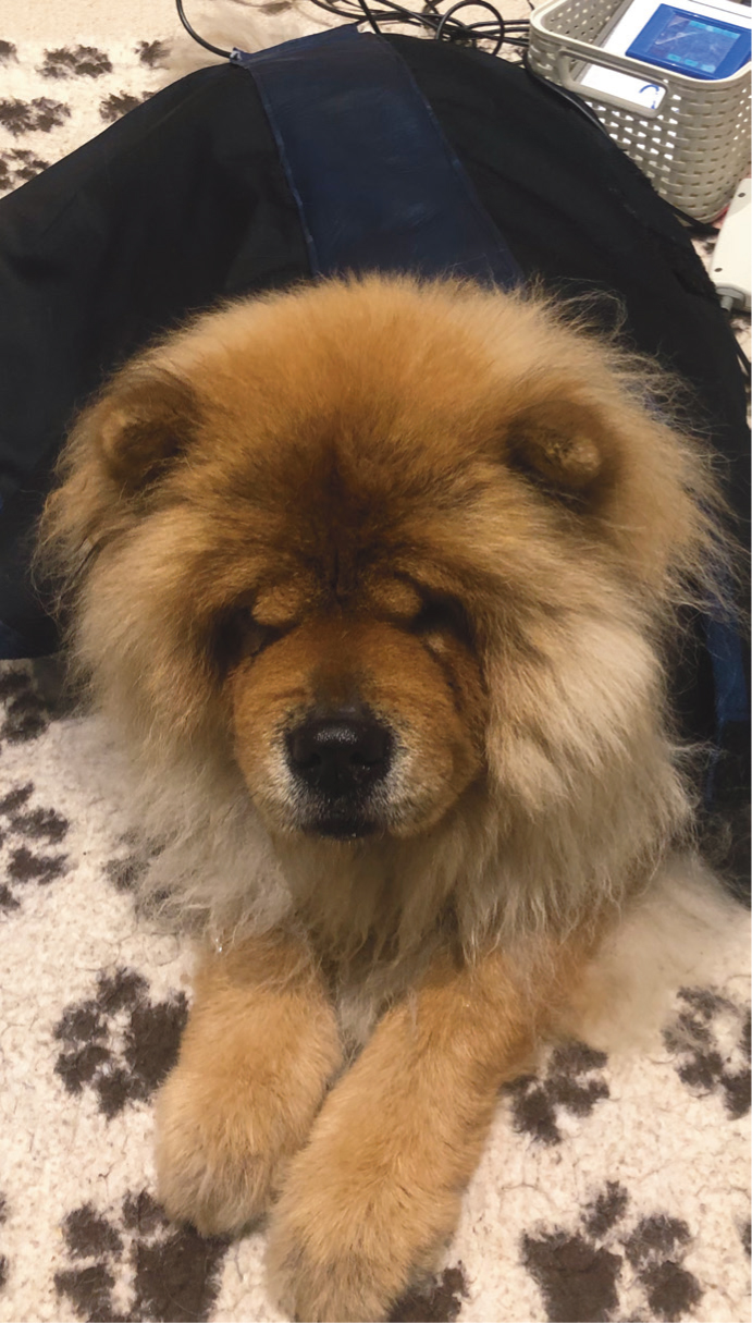
Photobiomodulation, which is a therapeutic laser (Figure 7), has become an increasingly popular treatment tool in veterinary medicine because of its potential to aid pain management, wound healing and the reduction of inflammation. The beneficial effects of photobiomodulation come from the the activation of cytochrome oxidase at the level of the mitochondria, which stimulates cell ATP production (Bruno et al, 2020). There is some evidence of axonal regeneration following spinal cord injury (Byrnes et al, 2005) and of improved neurological status following surgical treatment of dogs recovering from thoracolumbar disc extrusion (Bruno et al, 2020). However, the evidence is mixed and further research is required before definitive conclusions can be drawn.

Acupuncture
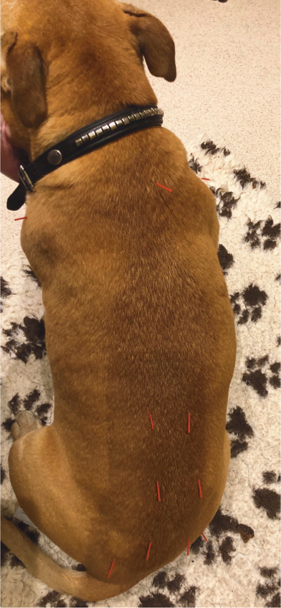
There is some evidence to suggest that acupuncture (Figure 8) can have neuroprotective effects for a patient following spinal injury, as a result of its effects on microglial activation and regulation of pro-inflammatory mediators, which can potentially exacerbate neural injury (Choi et al, 2010). Electroacupuncture has been demonstrated to enhance analgesia and accelerate motor function in patients suffering from intervertebral disc disease (Joaquim et al, 2010), as well improving pain caused as a result of various neurological conditions (Roynard et al, 2018). It is therefore an important adjunct to other physical rehabilitation therapies.
Conclusions
Most canine patients recovering from spinal cord injury will benefit from a multimodal approach to rehabilitation. This includes the use of physical therapies, such as physiotherapy and hydrotherapy, to assist with recovery of ambulation, muscle strengthening, coordination and enhancement of proprioception. Electrotherapy and acupuncture can also be used for pain control and to increase the potential for tissue regeneration and an improved neurological status. These therapies, when performed by suitably qualified individuals, together with pharmaceuticals for analgesia where required and high standards of nursing care, form an important part of patient care, as they can both facilitate recovery and improve patient welfare.
KEY POINTS
- A neurological assessment before initiation of rehabilitation therapy is key, to identify pain, neurological deficits and areas of weakness.
- Different approaches are used for ambulatory versus non-ambulatory patients.
- Early intervention with rehabilitation therapy has been shown to improve patients' return to function.
- A multimodal approach combining exercise therapy, electrotherapy and pain-relieving techniques is likely to achieve the best outcome for the patient.


