The shoulder is the most mobile joint of the canine appendicular skeleton. The majority of the available movement is within a sagittal place (flexion and extension), however there is also a degree of rotation, both internal and external, adduction and abduction (Marcellin-Little et al, 2007). This wide range of motion is afforded by the ball and socket nature of the joint, formed by the glenoid cavity and humeral head. Stability of the shoulder joint is offered by the tough, fibrous joint capsule. This comprises the medial and lateral glenohumeroid ligaments and tendons from large surrounding muscles, located inside or outside the joint capsule (Sidaway et al, 2004).
Clinical examination and diagnostics
The shoulder joint is comparatively challenging to examine thoroughly because of the bulk of soft tissue coverage surrounding it and the difficulty in isolating it as the source of pain without inadvertently stressing the elbow at the same time. During physical examination, the surrounding soft tissues should be evaluated for signs of discomfort and asymmetry, and the joint for available range and pain response during flexion and extension. It is also important to assess the shoulder for signs of instability. This is performed by assessing cranio-caudal and medio-lateral movement of the proximal humerus relative to the distal scapula (Marcellin-Little et al, 2007). The biceps tendon should also be assessed by palpation, and by extending the elbow with the shoulder in a flexed position (Bruce et al, 2000). Medial shoulder instability should be ruled in or out by assessing the level of available abduction, which is normally around 30° (Henderson et al, 2015).
During the examination, the patient should also be assessed for neck pain and issues with the elbow, carpus and digits to rule out alternative or co-existing issues which may present with similar clinical signs, such as lameness or pain on palpation.
Diagnostic imaging of the shoulder often begins with radiography, but may also require MRI, arthroscopy and/or ultrasonography to achieve a confirmed diagnosis (Henderson et al, 2015). These modalities are used to assess the shoulder for:
- Developmental issues, including osteochondrosis dissecans
- Traumatic injuries, including fractures and luxations
- Soft tissue injuries to the surrounding ligaments and tendons, including partial tears, chronic tenosynovitis, adhesions and contractures
- Osteoarthritis (Morris and Anderson, 2013).
Goals of rehabilitation
The best possible outcome following rehabilitation for the patient and owner is a full return to function and, where possible, this forms the end goal of any rehabilitation treatment programme. In order to achieve this, the process of rehabilitation must be broken down into stages. These stages in healing are similar regardless of the causal factor of the shoulder injury/condition. The differences between rehabilitation programmes for different conditions of the shoulder are the time frame through which the stages can be progressed and the likelihood of the patient achieving a full return to function.
Stage 1 — immobilisation and analgesia
The goals during the first stage of rehabilitation are to provide the patient with effective pain management and to allow tissue healing through immobilisation and rest. Maintaining range of motion in the joints and slowing down atrophy of the surrounding musculature are also desirable (Millis and Ciuperca, 2015).
Analgesia is oft en initiated with the use of non-steroidal anti-inflammatory drugs (NSAIDs) by the veterinary surgeon. However, during rehabilitation the therapist will oft en administer electrotherapy, such as pulsed electromagnetic field therapy and/or LASER, as these modalities have beneficial effects on both the healing process and on pain pathways (Millis and Cuiperca, 2015; Pryor and Millis, 2015).
Ice packing may also be beneficial to reduce the inflammatory cascade, in particular for the first 3 days following injury or surgery (Millis and Ciuperca, 2015).
Localised immobilisation of the shoulder joint (through use of a harness or splints) is desirable in some cases, despite the negative effects on joint range of motion. One example of this is in cases of medial shoulder instability (a stress injury to the medial glenohumeral ligament) whereby use of hobbles such as the ‘DogLeggs shoulder stabilisation system’ to prevent abduction and allow stabilisation of the shoulder joint is beneficial to facilitate recovery (Henderson et al, 2015). A system like this may also be beneficial to assist in the recovery of certain types of scapular fracture, and in cases of luxation or subluxation. Otherwise, if the shoulder is structurally sound, maintaining joint range of motion is beneficial to the patient in the long term (Saunders, 2007).
Resting the patient with shoulder pathology not only protects the patient from further injury, it also protects a surgical repair site from excessive stress, strain and potential mechanical failure, and allows tissue repair to take place (Dorn, 2017). This is achieved by limiting the activity of the patient to very short lead walks and preventing excessive force through jumping and landing. It may be facilitated by use of a crate in situations where the dog is boisterous or is unsupervised at certain times of day (Dorn, 2017). The duration of rest of this nature before gradual reintroduction to load bearing exercise is dependent on the nature of the pathology and can range from a few days for a mild sprain to 6 weeks or more for a fracture.
Range of motion during the first phase is best maintained using passive range of motion (PROM) exercises. This involves the therapist (and often the owner following demonstration of technique) moving the joints of the limb, with focus on the shoulder joint, passively through flexion and extension until natural resistance from the patient is met. This should be done in a slow and controlled fashion, while supporting the limb, for around ten repetitions (Marcellin-Little and Levine, 2015). This exercise should be completed 2–3 days daily and is shown in Figure 1. In addition to maintaining range of motion, PROM has a positive impact on the healing process, as it assists with nourishment of the cartilage, decreases oedema and adhesion formation, and assists with pain management (Marcellin-Little and Levine, 2015).
At this stage, slowing down muscle atrophy is mainly achieved through judicious use of pain relief to promote load bearing while the patient is moving around the home and during the very short, controlled lead walks, the benefit of which on recovery should not be underestimated (Saunders, 2007). As the buoyancy properties of water minimise impact on the joints, the underwater treadmill or swimming may be useful, again depending on the nature of the injury (Saunders, 2007).
Stage 2 — range of motion and strengthening
Once the rehabilitation therapist and veterinary surgeon have completed their assessment and are content that the patient's pain levels have stabilised and that they are able to load the limb, they can progress to the next stage of the rehabilitation process. The goal here is to open up the available range of motion in the shoulder joint and initiate low impact strengthening exercise to begin to build the surrounding musculature. Normally, it is advisable that the patient attend the rehabilitation clinic on a twice weekly basis, and an exercise programme consisting of some or all of the exercises below should be given to owners to complete twice daily at home.
Increasing joint range of motion is achieved through continuing PROM exercises as described above, supplemented with active range of motion exercises, three examples of which include use of cavaletti poles, use of a physio exercise ball and ‘high-five’ exercises, as well as stretches.
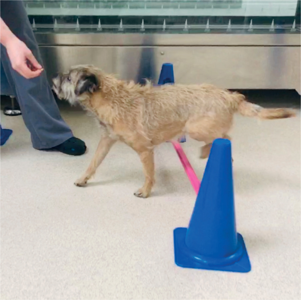
Cavaletti poles promote shoulder flexion (Figure 2) and are best performed with the dog wearing a harness which does not restrict shoulder movement, on-lead to enable slow, controlled movement. The height and spacing of the poles should be adjusted according to the size of the dog to promote exaggerated shoulder flexion (Marcelin-Little et al, 2007).
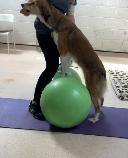
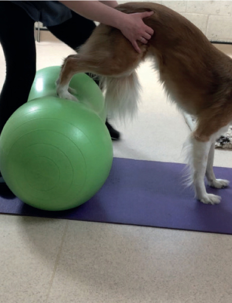
Therapeutic exercise balls — round or peanut shaped — are an excellent way to promote increased shoulder flexion (with the dog's forelimbs raised on the ball as shown in Figure 3) and extension (with the hindlimbs positioned on the ball as shown in Figure 4) (Marcelin-Little et al, 2007). The fore or hindlimbs are lifted on to the ball, which is held in a steady position, and the dog encouraged to stay (often with use of a treat) for 10 seconds or so at first, building gradually towards 30 seconds.
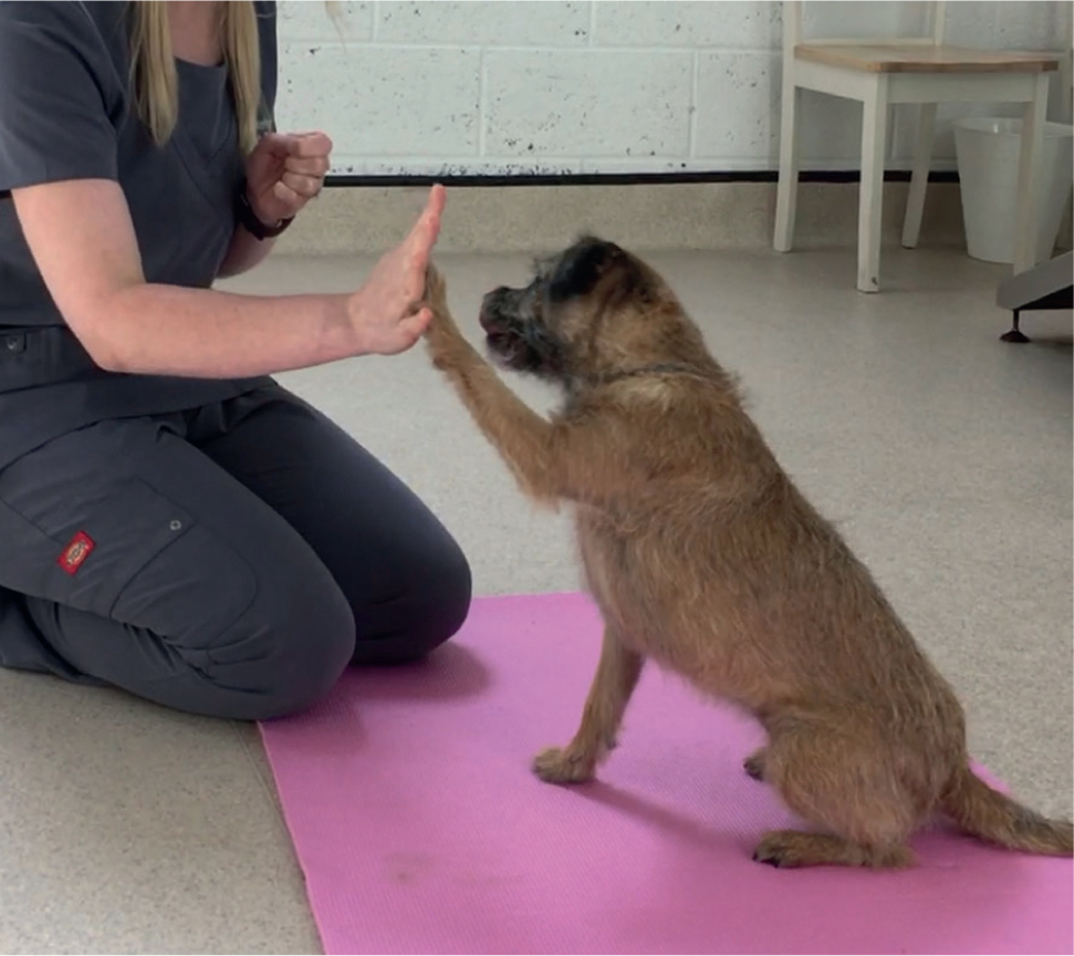
Asking the patient to bring the shoulder into extension to ‘high-five’ the therapist (Figure 5) is also an excellent and easily-performed active range of motion exercise (Prydie and Hewitt, 2015). This exercise is normally initiated with three repetitions, adding more as the patient progresses.
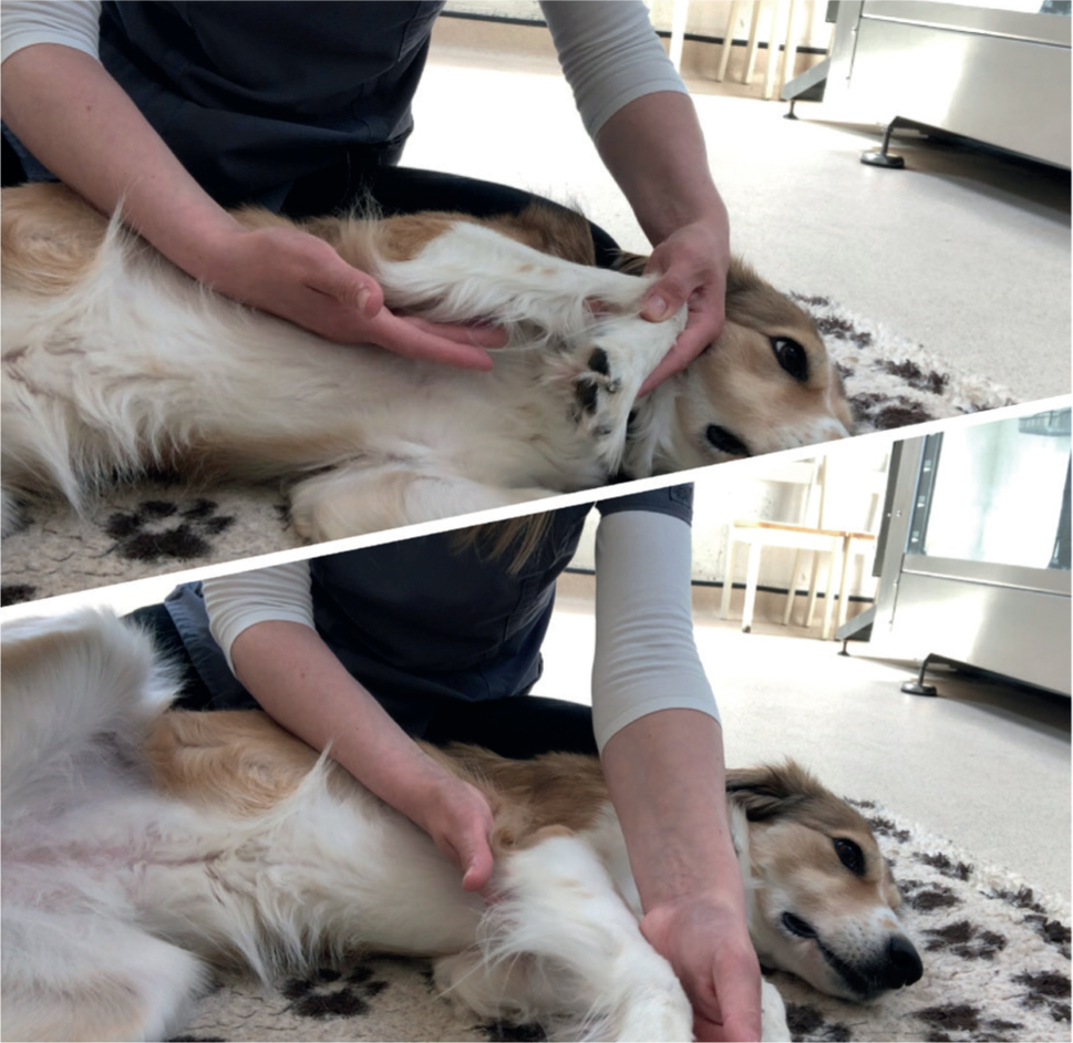
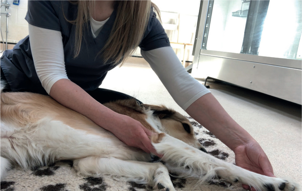
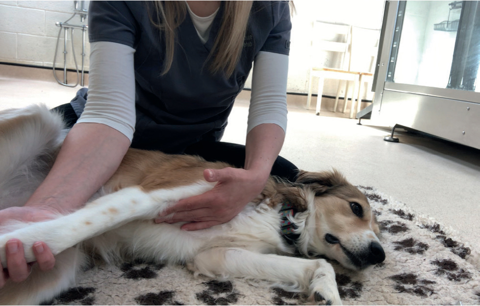
Stretching promotes elongation of the muscle tissue and, for the shoulder region, is best performed with the patient in lateral recumbency with the affected limb uppermost, after PROM exercises (Marcellin-Little and Levine, 2015). The shoulder is extended and gentle pressure applied to the caudal humerus while the limb is supported underneath by the therapist (Figure 6). The patient is monitored to ensure that they are comfortable, while the limb is held in this position for at least 10 seconds (Marcellin-Little and Levine, 2015). The limb is then retracted so the shoulder is in a flexed position and gentle pressure is applied to the cranial humerus, with the limb supported underneath (Figure 7), and held again for at least 10 seconds. This would be repeated three times.
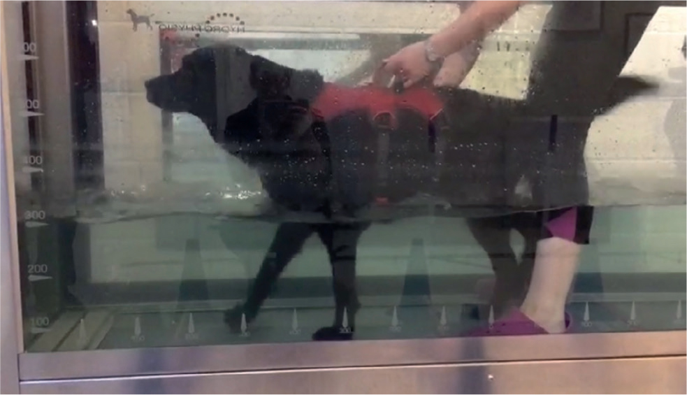
All of the active range of motion exercises described above promote muscle contraction as the patient performs the exercise, and so also serve as strengthening exercises in conjunction with the initiation of short (around ten minutes), frequent lead walks and use of hydrotherapy in the form of swimming or underwater treadmill walking (the optimum water height differs for different conditions of the shoulder (Figure 8) (Drum et al, 2015).
When return to a full range of motion (or, for long-term conditions, a significant improvement) has been achieved, along with reasonable muscle development, the patient can be advanced to the following stage.
Stage 3 — continued muscle strengthening
The next stage in rehabilitation is to increase the duration and intensity of the exercises to promote continued muscle strengthening and range of motion.
The passive and active range of motion exercises, plus the stretching exercises outlined above, should be continued. Walking should be increased by adding 5 minutes per week onto the length of each walk. Once the patient reaches around 30 minutes of walking without displaying signs of fatigue, this should include gentle downhill walks to increase weight bearing on the forelimbs (Doyle, 2004). The speed and duration of underwater treadmill walking, or the intensity of swimming sessions, should be increased gradually under professional supervision. At the end of this stage, the patient will have ideally recovered full range of motion of the shoulder joint and have even, well developed musculature between the forelimbs.
Stage 4 — return to function
The final stage in rehabilitation is to gradually reintroduce higher impact exercises that the patient had participated in before injury or surgery, such as running off-lead and playing with other dogs. For some ongoing conditions, such as osteoarthritis, certain activities may be best avoided in the long-term as part of a management strategy; but for conditions where full recovery is likely, gradual reintroduction of these activities should be initiated before winding down physical therapy so the patient can be signed off from treatment.
Conclusion
As always, rehabilitation should be supervised by suitably qualified staff (belonging to one of the following registers: RAMP, IRVAP, NAVP, ACPAT) to which the patient has been referred by a veterinary surgeon. An exercise prescription for use between sessions with the therapist, and owner commitment to administering these exercises, are integral to the success of any rehabilitation programme. When used in this way, physiotherapy and hydrotherapy can be a fundamental tool in the recovery from injury and surgery of canine patients with conditions affecting the shoulder, and in the long-term management of those with incurable developmental conditions or osteoarthritis.
KEY POINTS
- The shoulder is an extremely mobile joint, stabilised by a fibrous joint capsule and surrounding ligamentous and tendon structures.
- Assessing the shoulder joint can be difficult because of the overlying soft tissue structures and the ability to mobilise the joint without affecting the elbow.
- Injuries to the shoulder joint undergo four stages of rehabilitation regardless of the injury/condition.
- Rehabilitation therapies play a vital role in this, alongside pharmaceutical or surgical management where applicable.


