Elbow pathology is a common cause of forelimb lameness in the dog (Scott and Witte, 2011). Unlike the shoulder, which is affected by many conditions involving soft tissue structures providing stability, the majority of issues affecting the elbow arise from the bony constituents, including incongruities, dysplasia, osteochondrosis dissecans and osteoarthritis, as well as injuries resulting from trauma.
Examination and diagnosis
Clinical examination of an animal with forelimb lameness must consist of a thorough examination of all joints. Particular attention should be paid to the structures and stability of the shoulder joint, because of the prevalence of pathology in this region, as well as the elbow before concluding which areas warrant further investigation (Cook and Cook, 2009).
Evaluation of the elbow joint relies upon diagnostic imaging modalities, such as radiography using oblique positioning, to increase the likelihood of visualising loose fragments. Other modalities include computed tomography, magnetic resonance imaging and/or scintigraphy, although in some cases, arthroscopy for direct visualisation of the bony structures may be necessary to confirm diagnoses (Fitzpatrick et al, 2009).
Irrespective of the condition, several studies demonstrate the benefits of rehabilitation therapy to animals following injury or surgery, with the ultimate goal of returning them to their previous ability in a timely and safe manner (Canapp et al, 2009). As with the shoulder joint, described in the previous article in this series (10.12968/coan.2020.0042), the process of rehabilitating an animal with elbow pathology must be broken down into stages, beginning with an assessment by the therapist, before progressing through the next stages.
Stage 1: immobilisation and analgesia
The goals of stage 1 include:
- Analgesia (and reduction of inflammation)
- Tissue healing (through rest and immobilisation)
- Maintaining joint range of motion, where complete immobilisation is not necessary
- Minimising atrophy
- Encouraging early load bearing (Canapp et al, 2009).
Rehabilitation therapists have multiple modalities which can be used to encourage the animal's progress through stage 1, alongside medical management provided by the referring veterinary surgeon.
The reduction of inflammation can be supplemented through use of cryotherapy, which causes vasoconstriction, decreased motor and sensory nerve conduction and analgesia. Specific cryotherapy equipment is available to purchase but manual application of cold therapy can be used instead, by wrapping a wet swab around an icepack and placing it around the entire elbow for 15–20 minutes (Canapp, 2010).
Electrotherapy can also be advantageous in controlling pain. It generally includes pulsed magnetic field therapy and laser therapy. Photobiomodulation therapy, often referred to as low-level light therapy, has been shown to reduce pain scores in dogs suffering from osteoarthritis of the elbow, even reducing the need for non-steroidal anti-inflammatory drugs. The mechanism of action is through activation of enzymatic pathways within the mitochondria of the cell, which has positive influences on a number of processes including wound healing and analgesia (Looney et al, 2018).
Maintaining elbow range of motion is extremely important for function in dogs. Full extension of the elbow is required during gait and significant flexion is needed for everyday activities, such as using stairs or stepping over obstacles (Millis and Levine, 2014). This can be achieved with the use of passive range of motion exercises (discussed in prevously published article on rehabilitation of the shoulder) and joint mobilisations, discussed below. Early range of motion exercises nourish the articular cartilage and assist in the formation and structure of collagen tissue deposition. The primary goal of this type of exercise is to establish elbow extension and reduce the risk of contractures from the elbow being held in flexion (Canapp et al, 2009). The elbow is predisposed to flexion contractures as a result of the anatomy of the joint and surrounding structures. The congruency of the joint articulations, the tight nature of the joint capsule with a predisposition for developing adhesions at the cranial aspect and the position of the biceps/brachialis complex, which attaches to the joint capsule before crossing the joint, are all contributing factors (Canapp et al, 2009).
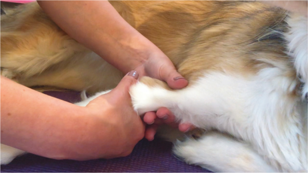
Joint mobilisations are passive movements which are oscillatory in nature. To encourage elbow extension, the therapist approaches the forelimb ventrally, stabilising the distal humerus with one hand, while the other hand applies gentle pressure to push the radius and ulna caudally, to extend the elbow to its end of available range (Figure 1), then back to midrange at a speed of around 3–4 repetitions per second, for 60 seconds. The range of motion should be assessed after each set and if there is an increase in available range, this exercise can be repeated up to three times within one treatment session (Millis and Levine, 2014).
Throughout stage 1 effective analgesia is of primary importance, to encourage the animal to load bear without the risk of aggravating pain. Therapeutic exercise can also be initiated, with the aim of minimising atrophy and encouraging early loading of the affected limb. Safe exercises to initiate at this stage include:
- Walking on a lead, starting with 5 minutes, three times daily, increasing to 20 minutes by the end of the first stage
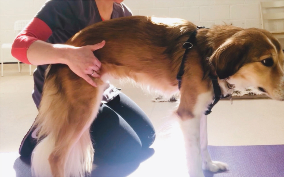
- Rhythmic stabilisations (Figure 2). While the patient is in a standing position, the therapist applies gentle pulses in either the inguinal region or at the level of the shoulders, to shift weight either in a cranio-caudal or medio-lateral direction, for around 30 seconds, three times daily
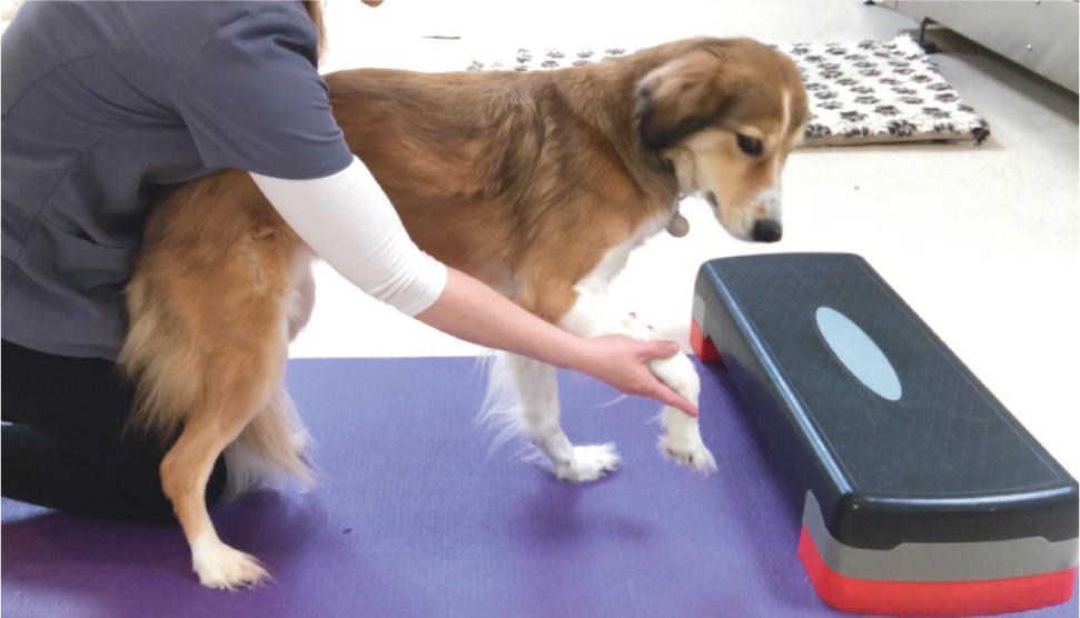
- Three-legged stand (Figure 3): The therapist lifts each of the patient's limbs from a standing position, to encourage strengthening of the limbs that remain standing by bearing the weight, alongside a gentle flexion of the elevated limb (Canapp, 2010).
Stage 2: improving range of motion and continued strengthening
Once pain levels are under control, any skin wounds or incisions have healed and lameness has improved (together with the go-ahead from the referring vet or surgeon), the focus of rehabilitation can progress to stage 2. This stage involves improving joint range of motion further and initiating strengthening to address inevitable atrophy.
As well as continuing passive range of motion exercises, stretching also facilitates an improvement of joint range of motion and helps prevent contractures (Marcellin-Little and Levine, 2015). When performing any stretching exercise, it both increases efficacy and safety to first warm the tissues using a heat pack applied to the desired area for 5–10 minutes. Stretching the elbow is best performed following passive range of motion exercises, with the limb supported underneath by the therapist. The limb is held in the position of extension where resistance is met, for at least 10 seconds, repeated three times daily. The anticipated gains in range of motion by stretching are between 5–10° per week acutely and 3–5° per week in more chronic cases (Marcellin-Little and Levine, 2015). This can be measured by goniometry, the normal range for which is 36–165° in the canine elbow (Marcellin-Little and Levine, 2015).
It is at this stage that hydrotherapy (either on an underwater treadmill or in a hydrotherapy pool with a fully qualified rehabilitation practitioner) is often initiated, alongside therapeutic exercises to help recovery. Hydrotherapy has been shown to increase range of motion of the elbow and forelimb stride length in one session alone, in both healthy patients and, to a greater degree, in those suffering from elbow dysplasia (Preston and Wills, 2018). This is hypothesised to be a result of both the buoyancy allowed by the water, which encourages the movement of joints into an improved range of motion and the thermotherapeutic effects of the warm temperature of the water (usually around 30 °C), which can improve muscle elasticity and joint extensibility, as well as offering analgesia (Preston and Wills, 2018). Hydrotherapy has the additional benefits of improving cardiovascular fitness, but as it pertains to the rehabilitation of the elbow joint, of strengthening musculature, with the provision of resistance by the water (Wild, 2017).
The following therapeutic exercises are examples of strengthening exercises which can be also be included at stage 2.
- Continuing the three types of strengthening exercises outlined in stage 1, but increasing their intensity. Specifically, increasing the walking time by 5 minutes per walk per week, the length of time and height of the elevated limb during the three-legged stand and by elevating the hindquarters of the animal on to a step, to increase load bearing through the forelimbs, while performing the rhythmical stabilisations
- Introducing cavaletti poles (discussed in the article on rehabilitation of the shoulder)
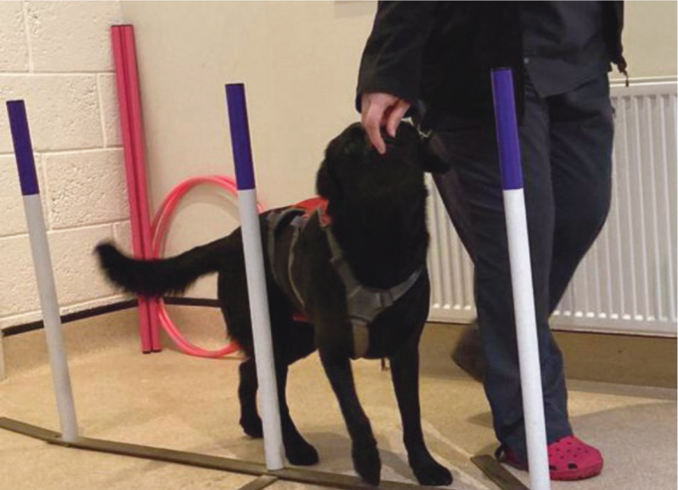
- Introducing weaving exercises (Figure 4), where the patient is led in and out through weave poles in a slow, controlled fashion, increasing load bearing through the inside limb, as the patient moves around the poles.
By the end of stage 2, it would be expected that the animal had a good, comfortable range of motion of the joint, together with a significant development in the musculature of the limb. This can be assessed objectively by goniometry, circumferential measurements of limb musculature, or by assessing gait using video footage.
Stage 3: continued strengthening
At this stage, the goal is to focus on continued development of the musculature to strengthen the limb to the point that, by the end of this stage, consideration can be given to introducing higher impact activities, which the animal had participated in before their injury or surgery.
The following exercises are examples of those which may be included at this stage, in order to improve the strengthening of the forelimb:
- Increasing the range of terrain, gradient, speed and time of on-lead walking
- Continued hydrotherapy with increases in the intensity of the programme, such as length of time swimming, speed and length of sets on the underwater treadmill
- Introducing a down-sit exercise (Figure 5), whereby the animal is asked to lie down, then rise into a sit position, starting with five repetitions 2–3 times daily and increasing this gradually as the animal's strength improves
- Using an unstable surface such as a wobble cushion (Figure 6), initially under one, then both front feet, improves the proximal strength of the forelimb and proprioception (Canapp, 2010).
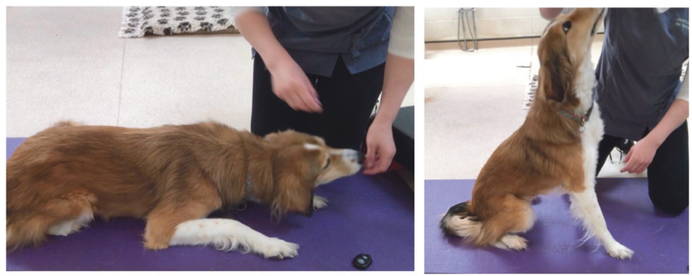
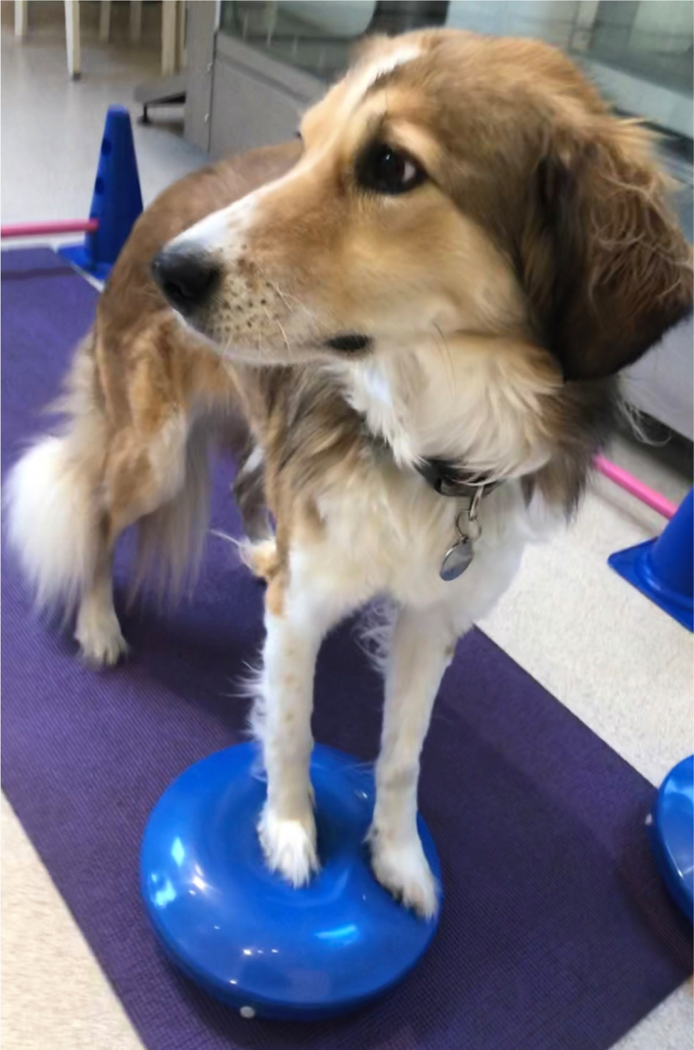
Many of the exercises described above, including walking on uneven surfaces, hydrotherapy, and the use of wobble cushions, have an additional benefit in that they enhance proprioception and neuromuscular control. For the canine patient, enhanced neuromuscular control means improved control of the elbow during exercise, by the surrounding musculature (Canapp et al, 2009).
Stage 4: return to function
If the intention is to return the animal to high impact activities, they must demonstrate the following upon examination:
- A complete and comfortable range of motion
- Even, well-developed musculature between the forequarters
- No evidence of pain
- No evidence of lameness on analysis of gait (Canapp et al, 2009).
For many dogs, where these factors cannot be met, it may prudent to discuss with the owner adaptations to their dog's lifestyle, to ensure they are well managed in the long term. This may include avoidance of certain activities, to avoid painful flare ups. For dogs where the above criteria can be met with satisfaction by the clinician, the first step is the gradual reintroduction of off-lead exercise, with monitoring to ensure there is no evidence of regression. If the dog copes well with this, gradual reintroduction to higher impact activities, such as playing with other dogs or ball play, can be initiated. Plyometric (jump) exercises, such as bounding through long grass or over raised cavaletti poles, encourage rebound action of the propulsive muscles (Hayes-Davies, 2014). This acts to further strengthen the limbs and prepare the dog for any higher impact sporting activities. Specific exercises for this can be selected and introduced to the rehabilitation sessions, depending upon the chosen activity.
Conclusion
With thorough assessments to ensure the canine patient is ready to progress through each stage, rehabilitating dogs with elbow disease or injury in this way can assist recovery and enable a full return to function, where possible. A combined approach, with communication between all members of the multidisciplinary team is essential, to ensure surgical, medical and rehabilitation applications are being used to complement each other. As always, it is essential to ensure any rehabilitation therapy is carried out by a suitably qualified individual (specific registers are available to ensure this).
KEY POINTS
- The majority of issues affecting the elbow arise from the bony constituents and include incongruities, dysplasia, osteochondrosis dissecans and osteoarthritis
- Several conditions affecting the elbow joint will benefit from rehabilitation therapies
- The stages of rehabilitation include immobilisation and analgesia, improving range of motion and strengthening, continued strengthening and return to function
- For many canine patients, rehabilitation will form part of the management strategy for long term conditions


