


Guinea pigs are popular pets in the UK, with estimated numbers of approximately 400 000, comprising 2% of the total pet population (Harrup and Rooney, 2020; Wills, 2020).
Osteoarthritis is commonly reported in many domestic animal species, but little information has been published regarding its prevalence in small mammal species (Redrobe, 2001; Keeble, 2006). Rabbits and guinea pigs have been used to develop osteoarthritis models in laboratory animal research (Arzi et al, 2012; Legrand et al, 2017). Reports suggest that the prevalence of this condition is high in small mammals, particularly in guinea pigs (Keeble et al, 2017).
What do we know about osteoarthritis in pet guinea pigs?
Although there have been significant advances in recent years in the field of pet rabbit medicine, pet guinea pig conditions are unfortunately less well documented and our understanding of their treatment and prevention is more limited (Meredith, 2015). In a study of pet rabbits in Finland, the prevalence of osteoarthritis affecting the distal limbs was found to be 2.4% with a significantly greater occurrence in older rabbits (>3 years old) (Mäkitaipale et al, 2015). This study also reported that 3.6% of rabbits were found to have facet joint osteoarthritis of the vertebral column.
By the age of 65, more than 80% of the human population has radiographic changes consistent with osteoarthritis in at least one joint site (Issa and Sharma, 2006). The case is similar in cats and dogs, where it is the most common arthropathy documented (Kuyinu et al, 2016).
A recent owner survey investigating the welfare of pet guinea pigs reported that 17% of guinea pigs sampled never stood on their hindlegs and 9% never showed ‘popcorning’ (jumping into the air, turning and running fast), which could easily be attributed to undiagnosed osteoarthritis (Harrup and Rooney, 2020). Of those that had been unwell, 2.4% were stiff when walking and 7.1% were lethargic, both of which are also signs that could be associated with pain and reduced range of joint movement caused by arthritis. Tibiofemoral osteoarthritis has been recognised as a significant health issue in pet guinea pigs in the Czech Republic (Minarikova et al, 2015). This study showed a prevalence of 5.6% for musculoskeletal disease in pet guinea pigs, with osteoarthritis being the most common musculoskeletal disorder identified (30/56 animals studied). A higher prevalence was recorded in older animals (above 2 years of age). One study by Keeble et al (2017) reports a radiographic prevalence of osteoarthritis in Guinea pigs of 100%, although the study group was small (n=23). This study also identified the stifle as the most commonly affected joint in guinea pigs. The true prevalence of osteoarthritis in the UK pet guinea pig population is currently unknown.
What causes osteoarthritis?
Osteoarthritis is a degenerative and inflammatory disease that is chronic and painful and affects the synovial joints, leading to reduced mobility. Osteoarthritis, or degenerative joint disease, is a disorder of movable joints, characterised by deterioration of articular cartilage, new bone formation at the articular surface (osteophytes) and subchondral bone involvement (Dequeker and Luyten, 2008). The condition is disabling and there is no definitive cure (Cucchiarini et al, 2016). Osteophytes represent new cartilage and bone development in osteoarthritic joints and arise from tissue in the chondro-synovial junction in response to mechanical joint injury, although their exact functional significance remains unclear (Sandell and Aigner, 2000). Osteophyte formation typically first appears on the medial tibial plateau, with associated hyperplasia of the synovial membranes, meniscal and femoral cartilage degeneration and subchondral sclerosis medially of the tibia and femur in some guinea pig lines as early as 9 months to one-year-old (Bendele et al, 1989) (Figure 1).
Primary (idiopathic) osteoarthritis has an inherited genetic background while secondary osteoarthritis is more common and occurs following a preceding problem or injury to a joint.
In guinea pig models, degenerative joint disease and osteoarthritis has been shown to spontaneously occur in older animals and most commonly affects the femorotibial joints (Turner et al, 2017). Adult weight is achieved between 8 and 12 months of age in guinea pigs, with an average life expectancy of 5–6 years. Bone development has been show to continue beyond one year of age in guinea pigs (Witkowska et al, 2014). It has been suggested that breed or strain differences contribute to the age of onset of osteoarthritis, with Dunkin-Hartley guinea pig strains being affected as early as 3 months old (Jimenez et al, 1997; Legrand et al, 2017). Histological changes noted on microscopy at postmortem include subchondral osteosclerosis, osteophytes, femoral condyle cysts, and ligament mineralisation (Turner et al, 2017). A guinea pig model for the study of human arthritis revealed that sedentary guinea pigs were more likely to develop severe osteoarthritis of the knee joints, partly because of their excess bodyweight and fat accumulation, but also as a result of functionally deficient joint tissue with poorer cartilage quality (Wallace et al, 2019).
Osteoarthritis may also occur secondary to dietary and husbandry factors in pet guinea pigs such as obesity, ulcerative pododermatitis, vitamin C deficiency or excess or inadequate exercise, improper substrate or joint trauma (Pignon and Mayer, 2021). Ovariectomy has also been shown to influence the development of osteoarthritis in female guinea pigs, with neutered animals being more likely to develop this disease (Yuan et al, 2017).
Low vitamin C levels have been associated with osteoarthritis, but even with vitamin C supplementation, osteoarthritis can still occur in guinea pigs, indicating that other factors are likely to be involved in the aetiology of this condition, as is recognised in other companion animal species (Minarikova et al, 2015).
What clinical signs may be seen?
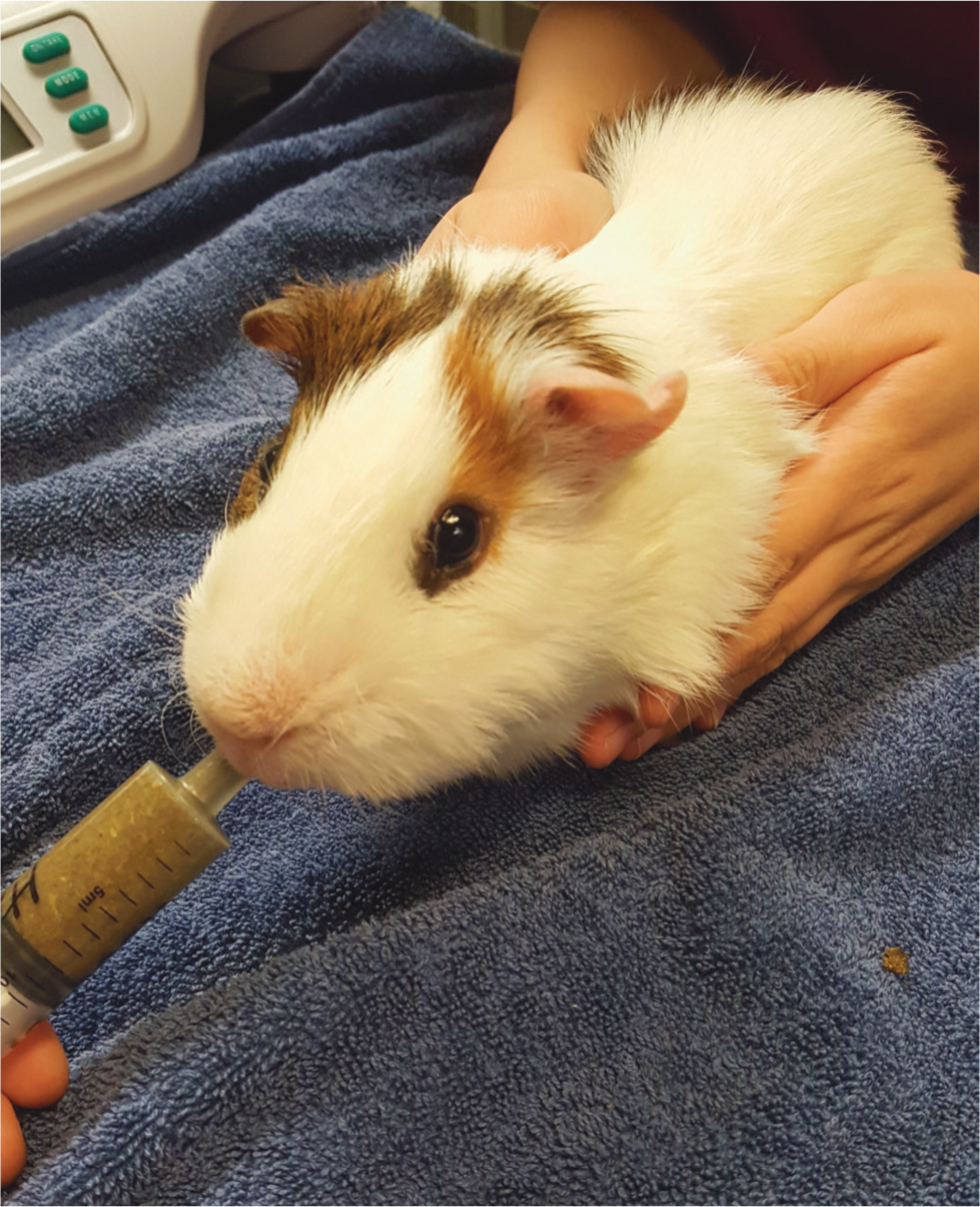
Osteoarthritis in guinea pigs often goes unnoticed by owners and veterinarians, even in advanced stages. Guinea pigs are not presented for routine health checks in the UK, unlike rabbits, which are routinely vaccinated. This poses a major welfare concern, particularly as arthritis can occur at a relatively early age in guinea pigs. The author recommends regular six-monthly health checks in all guinea pigs over three years of age. The examination should include manipulation of all joints to assess range of movement, particularly the stifles and hocks, as well as palpation of all joints for bone swellings and pain response. Affected animals often present for secondary reasons, such as weight loss, perineal faecal or urine staining, stranguria/dysuria and pododermatitis. Weight loss may occur secondary to chronic pain (Figure 2).
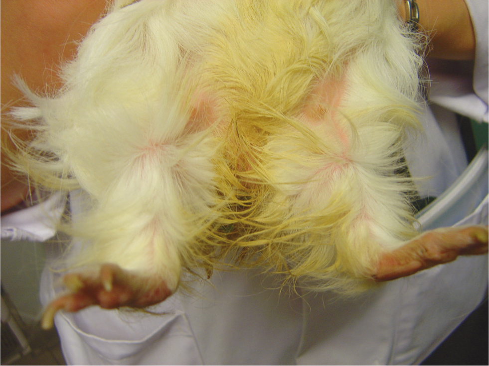
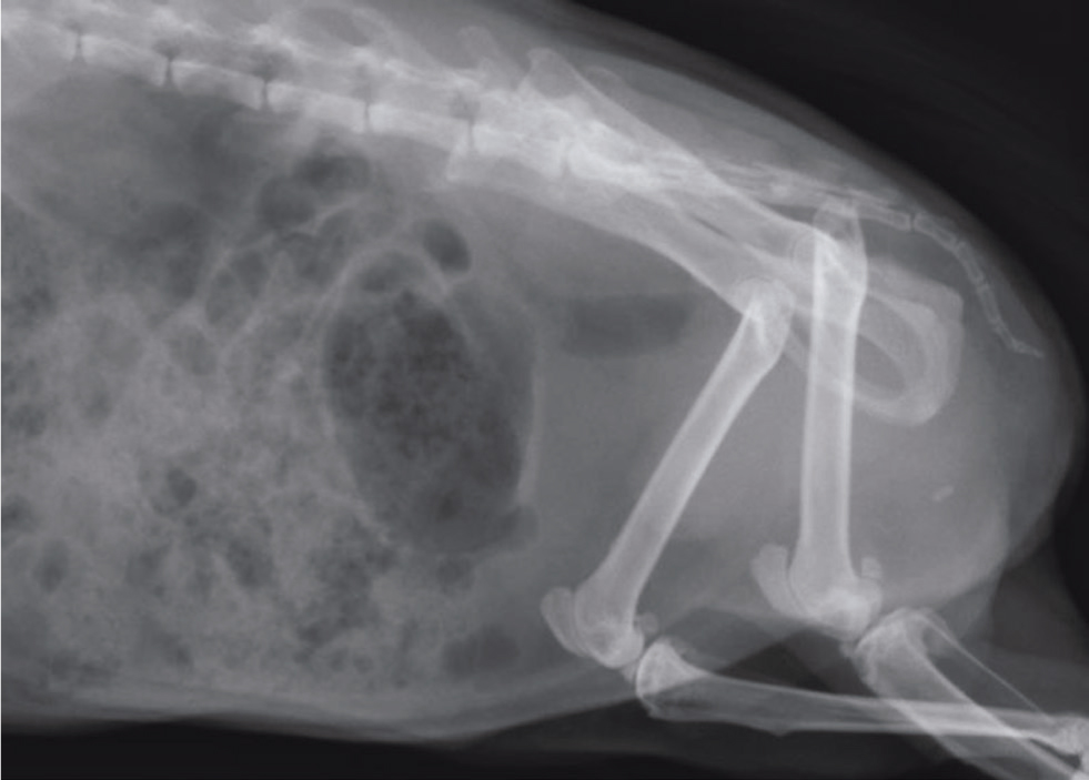
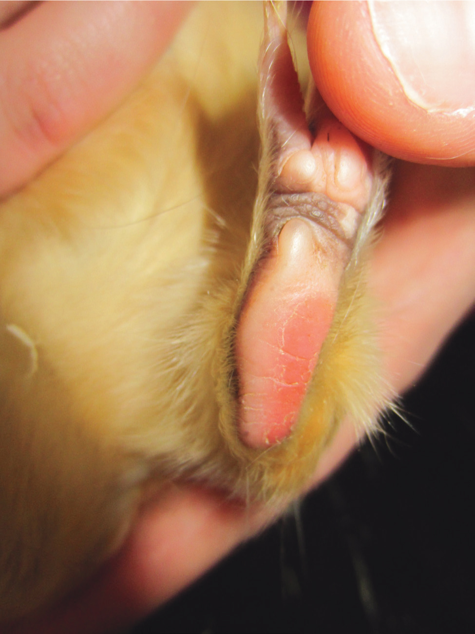
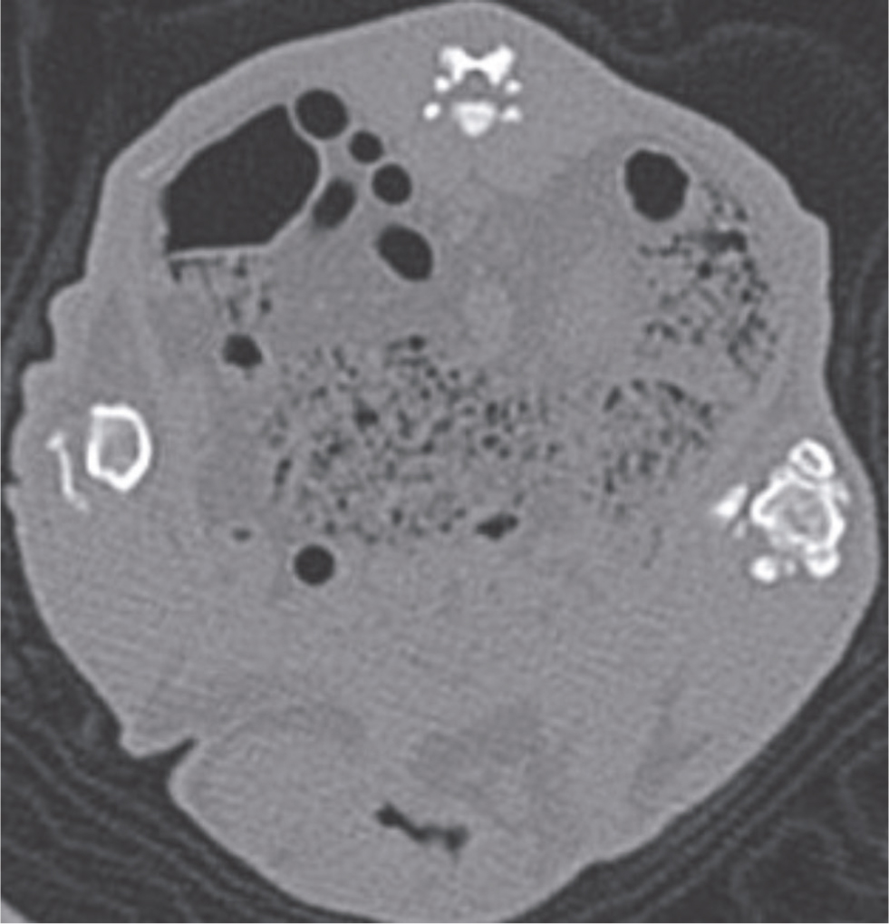
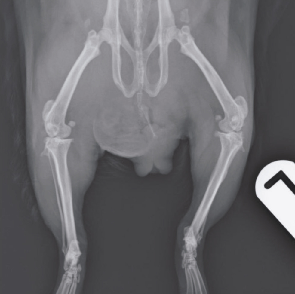
Inability to posture appropriately for urination or defecation can lead to perineal staining, secondary bacterial cystitis, hypercalciuria and urolith formation (Figures 3 and 4). Reluctance to walk and changes in weight bearing may result in pododermatitis, which can affect both the hind- and forelimbs, as well as nail overgrowth (Figure 5).
In advanced cases, animals may be reluctant to move or be visibly lame. Questioning the owner regarding changes in the animal's behaviour (increased lethargy or reduced ‘popcorning’ behaviour) as well as changes in gait and posture (reluctance to stand up on the hind limbs only) may also be helpful in assessing how affected the animal is by osteoarthritic joint changes.
How is osteoarthritis diagnosed?
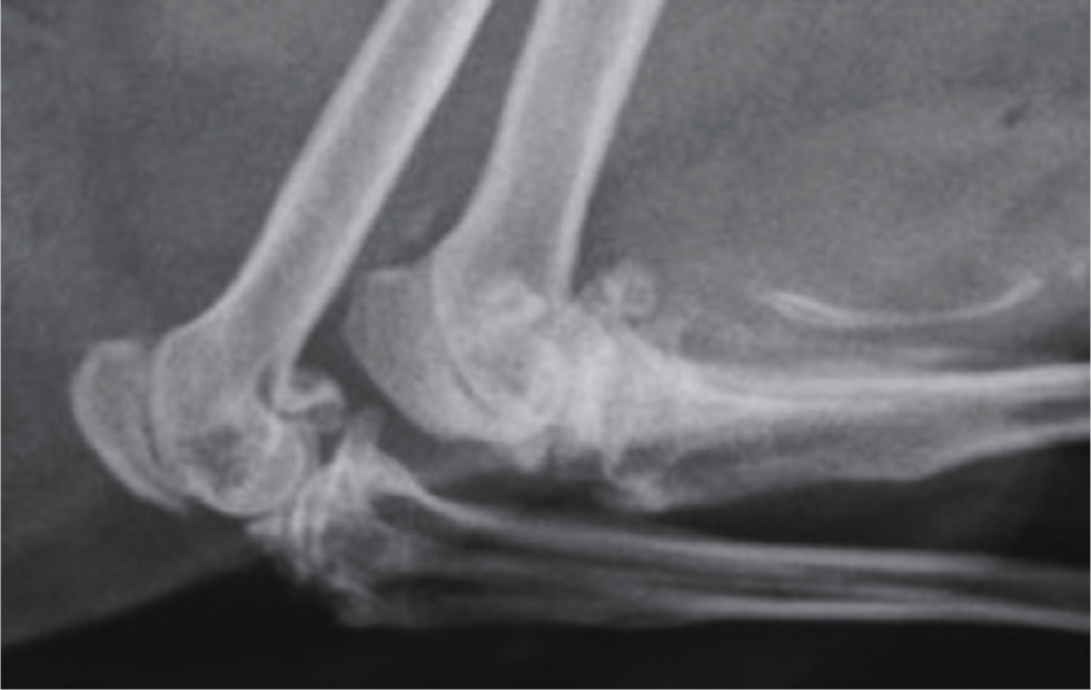
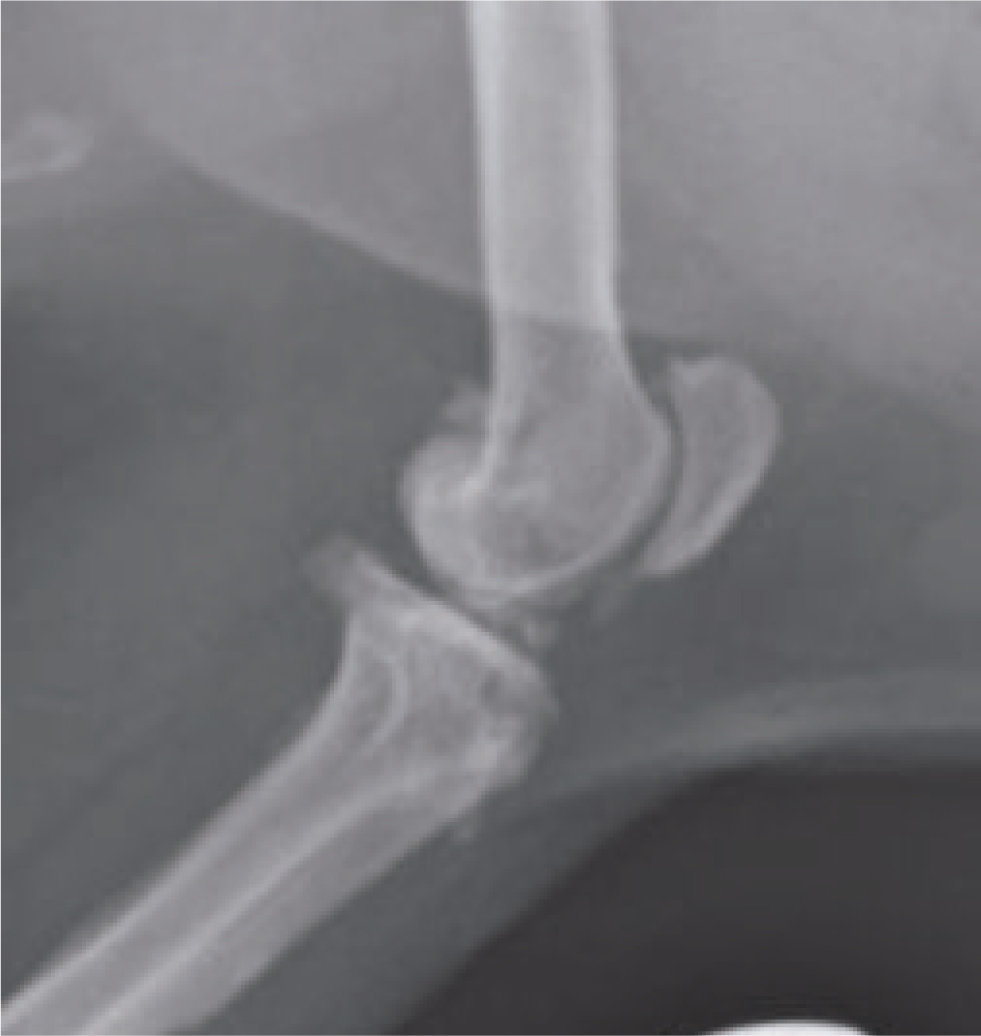
Although more advanced imaging techniques can be used to assess osteoarthritis, such as magnetic resonance imaging, computed tomography scan and arthroscopy (Burgkart et al, 2001; Lindblad, and Hedfors, 2005; Witkowska et al, 2014), conventional radiography is a standard and reliable imaging modality and is readily available to veterinary surgeons in practice (Chan et al, 1991) (Figure 6).
In human medicine, a radiographic grading system (K-L system) has been established, using a scale of 0 to 3, to evaluate the severity of this disease. This is based on evaluation of the number and size of osteophytes, the presence of joint space narrowing on weight bearing joints and the presence of sclerosis (Altman and Gold, 2007). Similar scoring systems have been adapted for companion animal species, but these rely on experienced personnel to interpret radiographic findings (Innes et al, 2004; Keeble et al, 2017). Keeble et al (2017) concluded that the stifle was the most commonly and severely affected joint in all species examined (rabbits, guinea pigs and chinchillas) radiographically. Common radiographic findings involving the stifle joint consisted of femoral osteophytes, tibial osteophytes, articular mineralisation, cranial meniscal calcification and caudal meniscal calcification, as well as patellar and fabellar osteophyte formation (Figures 7 and 8). This study reported a prevalence of 100% in guinea pigs, although the study group was small (n=23).
What treatments are available for this condition?
Treatment options in other species
Osteoarthritis management is well documented in other domestic species, particularly dogs. Sandersoln et al (2009) reviewed available literature to evaluate the efficacy of various therapies for the treatment/management of osteoarthritis in dogs. Treatments consisted of pharmacological agents, intra-articular agents, nutraceutical agents, alternative therapies, physical therapies, weight control and surgical techniques. The conclusion was that there was strong evidence that the non-steroidal anti-inflammatory drugs (NSAIDs) meloxicam, firocoxib, carprofen and etodolac were effective treatments, with moderate supporting evidence that glycosaminoglycan polysulphate, elk velvet antler, licofelone and food containing green-lipped mussel were effective in modifying structures involved in the progression of this disease in dogs.
In dogs, the mainstay of treatment is analgesia using NSAIDs, with carprofen, meloxicam, ketoprofen and firocoxib being clinically effective (Moreau et al, 2003; Autefage et al, 2011; Monteiro et al, 2019). These drugs can have side effects such as gastrointestinal signs and renal effects on platelet aggregation time, so lower doses have been recommended long-term in conjunction with tramadol in dogs to reduce these side effects (Monteiro et al, 2019). Other analgesics such as gabapentin or tramadol can be used in conjunction with NSAIDs.
The positive effects of various dietary supplements have been much debated in human and veterinary medicine, such as chondroitin sulphate, glucosamine, avocado-soya bean unsaponifiables, curcumin and polyunsaturated fatty acids (Comblain et al, 2016). Scientific data is needed to evaluate these products further and provide clear evidence that they work, rather than anecdotal evidence. More research is required regarding their bioavailability, particularly gastrointestinal absorption rates, pharmacokinetics and dosage regimen. Many have been shown to have anti-inflammatory and anti-catabolic effects.
Treatment options in pet guinea pigs
Treatment of osteoarthritis in guinea pigs is usually palliative, aiming to alleviate clinical signs, as cases often present in advanced stages with chronic joint changes already present. The mainstay of treatment is provision of appropriate analgesia long-term, but physiotherapy, soft bedding and weight reduction in obese animals should also be recommended (Pignon and Mayer, 2021) (Table 1). Product recommendation can be difficult as there are a wide variety of therapies available, often with little data existing regarding their pharmacokinetics and efficacy, with dosing often being extrapolated from other species (Lennox, 2010).
Table 1. Analgesic drug doses used in guinea pigs
| Analgesic drug | Dose rate, route and frequency |
|---|---|
| Buprenorphine | 0.05 mg/kg SC every 8–12 hours or 0.2 mg/kg every 5 hours oral or transmucosal |
| Butorphanol | 1–2 mg/kg SC every 4 hours |
| Carprofen | 2–5 mg/kg PO, SC, IM total daily dose give q12-24 hours |
| Gabapentin | 3–5 mg/kg PO every 12–24 hours |
| Ibuprofen | 10 mg/kg PO every 4 hours(Drug should only be used under direct veterinary supervision, following accurate dose calculations according to the animal's weight) |
| Ketoprofen | 1–2 mg/kg SC, IM every 12–24 hours |
| Meloxicam | 0.1–0.3 mg/kg PO, SC every 24 hours (This drug has been anecdotally used at higher doses up to 0.6 mg/kg PO every 12 hours by the author) |
| Morphine | 2–5 mg/kg SC IM every 4 hours |
| Tramadol | 5–20 mg/kg PO, SC every 12–24 hours (Efficacy and dosage in guinea pigs has yet to be established) |
IM = intramuscular PO = Per Os, SC = subcutaneous,
Adapted from Carpenter, 2018 and Quesenberry et al, 2021.
Oral supplements
Supplements containing growth cartilage components such as glucosamine and chondroitin sulphate may help reduce cartilage degeneration in guinea pigs (Taniguchi et al, 2012). These have been shown to protect against synovitis, inhibit degradation of cartilage and stimulate cartilage metabolism (McCarthy et al, 2007). In Taniguchi et al's (2012) study, supplements were started from three weeks of age and continued long-term (up to 18 months). Both supplement types showed an inhibitory effect on the progression of this disease when given orally at 200 mg/kg once a day. It should be noted that this study was carried out in a specific line of guinea pigs in an experimental situation and may not be directly extrapolated to the wider pet guinea pig population. It is not known whether this same effect would be seen in advanced stages of the disease, which is often when pet guinea pigs are presented to veterinarians. It is worth noting that clinical trials of dogs with diagnosed osteoarthritis, using a combination of these supplements orally for 70 days, showed significant improvements in pain scores, weight-bearing ability and disease severity, indicating that they may be beneficial even in advanced cases (McCarthy et al, 2007). Response to treatment was slower than in dogs receiving NSAID treatment with carprofen, so the use of both supplements and NSAIDs concurrently seems to be a sensible approach.
Physiotherapy
Physical therapy can be used in pet guinea pigs and is a common adjunctive treatment in pet dogs with osteoarthritis. A veterinary qualified physiotherapist should always be consulted before treatment is initiated.
Pulsed low-intensity ultrasound treatment for osteoarthritis has been described for both prevention and treatment of this condition in guinea pigs (Gurkan et al, 2010). This therapy was not found to be fully preventative but did reduce the severity of osteoarthritis and was found to be an effective treatment in affected animals, with better results in the early disease stages as opposed to the advanced stage. This could be a useful therapy in pet guinea pigs, although in this study animals were anaesthetised. Treatments were given for 20 minutes, for five days of each week.
Laser therapy and acupuncture have been described and evaluated in humans as adjunctive therapies for treatment of osteoarthritis and could potentially be beneficial in pet guinea pigs, although these modalities have not to date been fully evaluated in this species and their use in the treatment of osteoarthritis in the guinea pig remains controversial (Berman et al, 2004; Soleimanpour et al, 2014).
What lifestyle changes can be made to help?
Housing should be optimised for guinea pigs with osteoarthritis and this includes provision of non-slip, padded flooring. Avoiding stairs and ramps and lowering the litter tray on one side for easy access may help. Hayracks should be lowered so that they are easily accessible and do not require the animal to stand on its hind legs to reach the rack. Should they develop, secondary pressure sores should be monitored and treated as required. The perineum should be checked daily and cleaned if urine or faecal staining or faecal impaction occurs. Weight loss in obese animals and careful weight management is essential to alleviate pressure on diseased joints. Regular short periods of exercise are also important to prevent stiffness and build up muscle mass. Scatter feeding may encourage increased movement with foraging behaviour. The bedding area should be deep, soft and padded to avoid pressure sores and microwave heated pads or bags can be used, particularly in unheated or cold houses at night and during the winter months. Flooring such as laminate or tiles can be difficult for arthritic guinea pigs to walk on, so matting should be used to provide better grip, for example bath mats, yoga mats, carpet off-cuts, fleece vet bed and foam matting (Figure 9). Nail overgrowth is common in arthritic animals so it is important to keep guinea pigs' nails short with regular clipping.
Quality of life and animal welfare should be regularly assessed as this condition is painful, progressive and incurable. Sadly, euthanasia may be the kindest option in very advanced cases where there is chronic pain. These animals often lose weight and regular weight monitoring is useful in these cases.
Can this condition be prevented?
There is scientific evidence that glucosamine and chondroitin sulphate oral supplements are protective and can prevent against the development of osteoarthritis in guinea pigs when administered from an early age (Taniguchi et al, 2012). Ultrasound treatment could also be considered in the early stages of disease to prevent severe osteoarthritis from developing (Gurkan et al, 2010). Guinea pigs have been used as an animal model of osteoarthritis when investigating the protective actions of microbiota and vitamin C in preventing development of osteoarthritis lesions. Lyophilised inactivated culture and lipoprotein extract of Bifidobacterium longum were used with or without Vitamin C supplements and were found to act against dysbiosis during disease development, with vitamin C enhancing the effect (Henrotin et al, 2017).
Conclusions
Osteoarthritis is a chronic and debilitating condition. Commonly seen in advanced stages in pet guinea pigs, it is often overlooked by owners until it has become severe. Diagnosis is straightforward and is based on clinical evaluation and radiographic findings. Treatment is palliative with analgesia and lifestyle changes, although promising results have also been reported using supplements containing growth cartilage components in guinea pigs and these should be considered as adjunctive therapies. There is no cure for osteoarthritis and the disease is progressive. In advanced stages animal welfare may be compromised and euthanasia should be considered.
Guinea pigs have been extensively used as experimental models for osteoarthritis and our knowledge of pet guinea pig diagnosis, treatment and prevention for this condition is extrapolated from this research. More research is required into the incidence and development of this condition in pet guinea pigs. Greater awareness of this disease in pet guinea pigs among veterinary personnel and owners is essential for the early identification and treatment of this debilitating condition. More data are required on the pharmacokinetics, efficacy, therapeutic levels and dosage of analgesic drugs in guinea pigs to enable adequate pain relief to be given for this condition.
KEY POINTS:
- The true prevalence of osteoarthritis in pet guinea pigs in the UK is currently unknown but is thought to be high, especially in older animals.
- Clinical signs may be subtle in the early stages and are often missed by owners and vets, especially as pet guinea pigs are not routinely brought to veterinary practices for health examinations.
- Diagnosis, as in other species, is based on clinical and radiographic findings.
- Treatment is palliative and should be based around long-term analgesia, husbandry changes, weight reduction and physiotherapy.
- Dietary supplements, such as glucosamine and chondroitin sulphate, may be beneficial in the prevention and treatment of this disease in pet guinea pigs if given early on in the disease course.