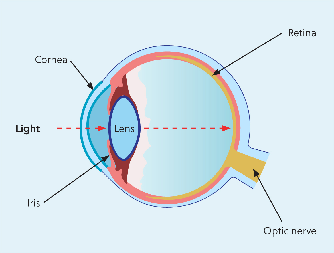
The ocular lens continues to grow throughout life, with new lens fibres forming around the equator like the rings of a tree (Ofri, 2018). The lens nucleus is at the centre of the lens, surrounded by the cortex and the acellular lens capsule. The new lens fibres (cortical fibres) are laid down on top of older lens fibres, gradually condensing them towards the centre of the lens (Figures 1 and 2). Nuclear sclerosis is a change in the lens consistently found in older dogs (over 7 years old) (Leiva and Pena, 2021), where the centre of the lens becomes compressed and hardened. The owners will sometimes have noticed a cloudy appearance to the lens, but this is not a cataract. In humans, nuclear sclerosis becomes evident in middle age, when suddenly reading glasses are suddenly required to read small print.


During fetal lens development, the lens has a blood supply, but these blood vessels regress and atrophy shortly after birth, resulting the the lens becoming almost entirely dependent on the aqueous humour for its nutrition (Leiva and Pena, 2021). The lens is optically transparent because of an absence of blood vessels and the highly ordered arrangement of lens proteins, which are mainly soluble crystalline proteins (Leiva and Pena, 2021). When these proteins are disrupted, this shows as an opacity of the lens called a cataract.
The eye is a site of immune privilege due to the blood-ocular barrier and dynamic immunoregulatory processes, which help to inhibit an inflammatory response (Miller, 2013). This helps to prevent damage to the delicate intraocular structures in the event of inflammation, which could result in blindness. The lens proteins have never been exposed to the ocular immune system, because the lens capsule surrounds the lens throughout its embryological development. The lens capsule allows diffusion of nutrients and waste, but not the passage of cells. If lens proteins leak out of the capsule, they are considered foreign proteins and a marked inflammatory response ensues, called lens induced uveitis (Ofri, 2018). This can be a consequence of either traumatic damage to the lens allowing lens material to escape from the capsule (phacoclastic uveitis) or secondary to a mature cataract when lens proteins start to liquefy and diffuse through the capsule (phacolytic uveitis) (Ofri, 2018).
What is the purpose of the lens?
The lens is responsible for fine-tuning focused vision, converging incoming parallel rays of light onto the retina. The focusing strength of a lens is measured in dioptres, and this varies between species. Each 1 dioptre can focus a beam of parallel light to a point within 1 metre. The cornea is the strongest lens in the eye, and is responsible for most of the refraction of incoming light (Miller, 2018). The lens also absorbs ultraviolet rays, protecting the retina from the harmful effects of this radiation (Augustin, 2014).
In mammals, the lens is suspended in the eye by zonular fibres, which extend from the ciliary body processes to the lens capsule at the equator of the lens (Samuelson, 2007). In humans and primates, when the ciliary body muscles contract, the zonular fibres relax, allowing the lens to form a more spherical shape, thereby increasing its refractive power (a process called accommodation) (Ofri, 2018). In carnivores, contraction of the ciliary muscles moves the lens anteriorly, allowing closer objects to come into focus. Posteriorly, the lens is in contact with the jelly-like vitreous, sitting in a depression of the vitreous face.
Classification of canine cataracts: three ways to classify cataracts
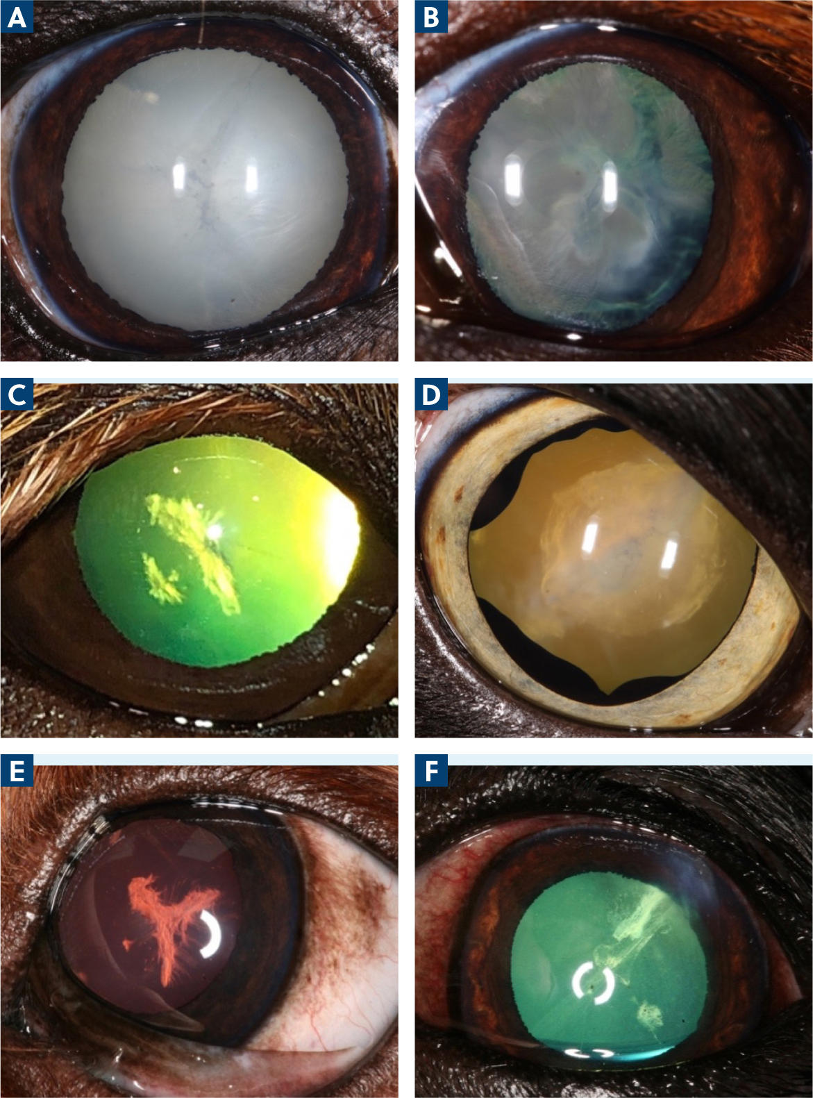
Cataracts can be classified according to how much of the lens is involved (incipient, immature, mature and hypermature) (Figure 3). An incipient cataract involves less than 15% of the lens volume, there is still a good tapetal reflex and detailed examination of the fundus is possible. Immature cataracts are between incipient and mature, with a reduced or hazy view of the fundus. Mature cataracts involve the whole lens, and the view of the fundus is obscured. Hypermature cataracts have advanced proteolysis of lens proteins, with reduced lens volume and wrinkling of lens capsule. Lens induced uveitis is common with hypermature cataracts, and there may be irreversible changes present, such as synechiae. As the lens volume shrinks, the lysed cortex may sink ventrally, resulting in a Morganian cataract.

Cataracts can be classified by their position; cortical or nuclear, anterior or posterior, axial or equatorial, and combinations. Last, cataracts can be classified by age of onset (congenital, juvenile, adult, senile). Congenital cataracts are present at birth because of disruption in development of the lens in utero. Juvenile cataracts develop shortly after birth and senile cataracts are common in geriatric dogs and tend to be cortical spoke-like opacities.
Diseases of the lens
Diseases of the lens present as changes in transparency, size, shape or position of the lens withiƒn the eye (examples include cataract, microphakia, lenticonus and lens luxation). The term ‘cataract’ encompasses any opacity of the lens or lens capsule, and are a common cause of blindness in dogs (Mellersh et al, 2009).
Cataracts can be congenital or acquired, and there are many causes including hereditary cataract, senile cataract, cataracts associated with intraocular inflammation, metabolic diseases, nutritional deficiencies, toxicities, trauma and as primary inherited defects.
Causes of cataracts
Hereditary cataracts
Heredity is a common cause of cataracts in purebred dogs, with over 100 breeds thought to affected with primary (inherited) forms of cataract (Mellersh et al, 2009). Each breed has a typical age of onset and location of the initial cataract, which may be static or progressive. The Labrador, for example, has a typical posterior polar subcapsular cataract that will often remain static and visually insignificant, but occasionally can progress to maturity, which causes blindness (Kraijer-Huver et al, 2008). There are many genes associated with hereditary cataracts in humans, and research by Mellersh et al (2006) identified a mutation in the HSF4 gene in three dog breeds affected by hereditary cataracts (Staffordshire Bull terriers, Boston terriers and Australian Shepherds). Not all hereditary cataracts will progress to cause visual impairment, but there is potential for hereditary cataracts to cause blindness which is why it is not recommended to breed from dogs with hereditary cataracts. Surgery is currently the only treatment available to remove cataracts and restore vision, and prevention is always preferable. Early detection is important to prevent breeding with dogs affected by hereditary cataracts, so it is advised to have the eyes examined by a qualified eye scoring panellist prior to breeding. Gene testing provides a simple diagnostic test to control disease in the affected breeds where gene tests are available, and this in combination with ophthalmic examination will help reduce hereditary cataracts and lead to healthier populations.
Organisations such as the European College of Veterinary Ophthalmologists and the British Veterinary Association, in association with the Kennel Club and International Sheepdog Society, keep a list of the breeds that are currently confirmed to be affected by breed-specific hereditary cataracts (Table 1). These lists are updated regularly as other breeds under investigation become confirmed. The results for Kennel Club registered dogs can be found on the Kennel Club website under health-test-results-finder (https://www.thekennelclub.org.uk/search/health-test-results-finder/).
Table 1. Inherited cataract in different dog breeds
| Breed | Inherited cataracts |
|---|---|
| Alaskan Malamute | Hereditary cataracts |
| Australian Shepherd | *Hereditary cataracts |
| Belgian Shepherd Dog | Hereditary cataracts |
| Bichon Frise | Hereditary cataracts |
| Boston Terrier | *Two forms – early and late onset |
| Cavalier King Charles Spaniel | Hereditary cataracts |
| French Bulldog | *Hereditary cataracts |
| German Shepherd | Hereditary cataracts |
| Giant Schnauzer | Hereditary cataracts |
| Irish Red and White Setter | Hereditary cataracts |
| Large Munsterlander | Hereditary cataracts |
| Leonburger | Hereditary cataracts |
| Miniature Schnauzer | Congenital hereditary cataracts, hereditary cataracts |
| Norwegian Buhund | Hereditary cataracts |
| Old English Sheepdog | Hereditary cataracts |
| Poodle (standard) | Hereditary cataracts |
| Retriever (Chesapeake Bay) | Hereditary cataracts |
| Retriever (Golden) | Hereditary cataracts |
| Retriever (Labrador) | Hereditary cataracts |
| Siberian Husky | Hereditary cataracts |
| Spaniel (American Cocker) | Hereditary cataracts |
| Spaniel (Welsh Springer) | Hereditary cataracts |
| Staffordshire Bull Terrier | Persistent hyperplastic primary vitreous, *hereditary cataracts |
Cataracts secondary to ocular disease and systemic disease
The second most prevalent cause of cataracts in dogs is diabetes mellitus. Diabetes mellitus leads to cataract formation in 70–80% of dogs within a year of diagnosis (Beam, 1999). The increased levels of glucose in the blood leads to raised glucose concentration in the lens, and the usual anaerobic metabolism of glucose by the hexokinase pathways becomes saturated (Leiva and Pena, 2021). Glucose metabolism is shunted to an alternative pathway using enzyme aldose reductase to reduce glucose to sorbitol. Accumulation of sorbitol raises the osmotic forces in the lens, leading to absorption of water from the aqueous humour which then causes lens fibre swelling, water clefting and intumescence (Ofri, 2018). Trials looking at topical aldose reductase inhibitors to delay onset of cataracts have shown some promise, but this product is not yet available in the UK (Kador et al, 2010; Balestri, et al 2022). Cats appear very resistant to cataract formation with diabetes mellitus, which is thought to be due to lower aldose reductase activity in this species compared to dogs (Richter et al, 2002).
Diabetic cataracts often progress quickly, with owners some-times reporting sudden onset of blindness over days to weeks. The swelling of the lens can be observed with slit lamp biomicroscopy showing a shallow anterior chamber and intumescence of the lens. Ocular ultrasound can also be used to demonstrate a swollen lens. A dog's lens should be no more than one-third of the axial length of the globe (approximately 7 mm in depth) (Samuelson, 2007). The intumescent lens is at risk of splitting the capsule as it swells, which can lead to loss of lens material into the vitreous. This results in intense lens-induced uveitis – called phacoclastic uveitis (van der Woerdt, 2000; Leiva and Pena, 2021). Eyes with pre-existing uveitis have an increased risk of complications after surgery (Gelatt and Wilkie, 2011) so it is recommended to perform diabetic cataract surgery as early as possible, as long as the patient is clinically well enough for general anaesthesia.
Frequently, cataracts occur secondary to ocular disease such as chronic uveitis (Ofri, 2018). Chronic uveitis leads to cataract formation because of impaired lens nutrition, which happens as a result of changes in aqueous humour composition. When a cataract and uveitis are seen together, a deduction as to which came first must be made, as cataracts can induce uveitis. Uveitis can also lead to posterior synechiae and/or iris rests (pigment deposits on the anterior lens capsule), causing capsular opacities (Hendrix, 2021). Trauma to the eye, both blunt and penetrating, can cause a cataract. Once the lens capsule is broached, the main concern is the development of a severe lens-induced uveitis (phacoclastic uveitis) that can be catastrophic for the eye. In cats, there is the additional concern of malignant sarcoma formation, which is sometimes seen after lens damage in this species (Wood and Scott, 2019).
Congenital forms of pigment spots on the anterior lens capsule are called persistent pupillary membranes, where remnants of the fetal blood supply to the lens remain. Persistent pupillary membranes can also present as strands of pigmented tissue originating from the iris collarette to either the iris, lens or the cornea (Hendrix, 2021).
On the posterior aspect of the lens, a congenital condition called persistent hyaloid artery and persistent hyperplastic tunica vasculosa lentis/persistent hyperplastic primary vitreous can cause cataracts when the remnants of fetal bloods supply remain in contact with the posterior capsule (Leiva and Pena, 2021). This can result in a wide range of opacities, from a single dot on the posterior lens capsule to a fibrovascular plaque or actual bleeding into the lens if the hyaloid artery remains patent. Certain breeds such as the Doberman and Staffordshire bull terrier are prone to this, and Staffordshire bull terriers are listed on the British Veterinary Association Eye Scoring Scheme as having confirmed hereditary eye disease.
Progressive retinal atrophy – a hereditary disease causing early retinal atrophy and blindness in many breeds – is often associated with cataracts due to oxidative damage caused by glutamate released by dying photoreceptors (Zigler et al, 1983; Leiva and Pena, 2021). The cataract typically starts in the posterior cortex, closest to the oxidants in the vitreous.
Other systemic conditions that induce cataracts include hypocalcaemia, toxins and nutritional causes (such as chronic vitamin E deficiency or milk replacers in the first week of life) (Leiva and Pena, 2021). Hypocalcaemia can be found in dogs with chronic kidney disease or hypoparathyroidism (primary and secondary). In puppies which have been hand reared on milk formula from an early age, a specific cataract with a perinuclear opacity and feathering at the suture lines has been reported (Ranz et al, 2002). Deficiency of amino acid arginine (and other amino acids) is the proposed cause.
Developmental cataracts
These are present at birth and can be either hereditary or as a result of an infection or toxic event during embryonic development of the lens. They may also be associated with other ocular developsmental conditions such as microphthalmia, multiple ocular anomalies, persistant pupillary membranes and persistent hyperplastic tunica vasculosa lentis/persistent hyperplastic primary vitreous.
Treatment of cataracts
Incipient cataracts have no effect on vision and treatment is not warranted. Immature cataracts can have mild to moderate effects on vision, and treatment is only recommended if the cataract is likely to progress. Mature and hyper mature cataracts cause marked visual impairment and tend to cause phacolytic uveitis (van der Woerdt, 2000), which (if left untreated) can result in longer term secondary glaucoma (Leiva and Pena, 2021). Treatment of phacolytic uveitis involves topical anti-inflammatory drugs such as steroids or non-steroidal anti-inflammatories. Cataract surgery is recommended once the eye is quiet (inflammation controlled), as surgeries on inflamed eyes have higher complication rates, and eyes where irreversible changes such as posterior synechiae have already occurred carry lower success rates and may be deemed unsuitable for cataract surgery.
Cataract surgery
Patient selection is important for the success of cataract surgery as not all eyes (and not all patients) are deemed good candidates for surgery. Early referral is key, so the eyes and the patient can be assessed and the risks versus benefits can be fully discussed with the owner. Incipient cataracts have an insignificant effect on vision, so treatment is not warranted, but monitoring for progression might be indicated.
Where irreversible changes such as posterior synechiae or preiridial fibrinous membranes have already occurred, success rates are likely to be lower (Gelatt and Wilkie, 2011). In cases such as acute phacoclastic uveitis, or where a foreign body has penetrated the lens, it may not be appropriate to delay surgery and this decision should be made after an ophthalmic examination and advice from a veterinary ophthalmologist. Surgery on immature cataracts prior to lens-induced uveitis tends to more successful (Gelatt and Wilkie, 2011) so early referral is recommended for the best outcomes. As part of the pre-operative assessment, it is recommended to screen for progressive retinal atrophy, especially if there is a history of nyctalopia and in predisposed breeds such as miniature poodles and Cocker spaniels. If the retina cannot be examined directly (due to maturity of cataract) then retinal function can be assessed with electroretinography. Ocular ultrasound can also be performed to check for retinal detachments, measure the size of the lens and assess for evidence of lens rupture.
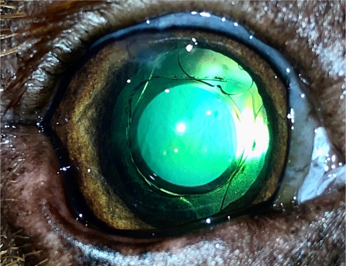
Cataract surgery (Figure 4) is performed using phacoemulsification, through small corneal incisions made just inside the limbus. Phacoemulsification was first developed in 1967 (Leffler et al, 2020) and describes using an ultrasonic handpiece to emulsify and aspirate the cataractous lens. Viscoelastic is used to maintain a formed anterior chamber, and capsulotomy (making a circular hole in the anterior lens capsule) is performed. The lens nucleus is removed with phacoemulsification and the lens cortex is removed with irrigation and aspiration. A prosthetic intraocular lens is then placed in the capsular bag if the bag is intact. Sometimes it is not possible to place an intraocular lens – for example, if the lens capsule is torn or unstable – and in these cases the eye can be left aphakic or a sulcus sutured lense can be placed (Gelatt and Wilkie, 2011). An aphakic eye is very long sighted, but dogs seem to cope well visually and certainly better than with the cataract.

Cataract surgery is more involved in dogs compared to humans for several reasons. Firstly, general anaesthesia is required in dogs, and often a neuromuscular block is administered to centralise the eye, which necessitates mechanical ventilation. Secondly, post-operative inflammation is more marked in dogs, partly because the cataracts are usually more advanced and the lens is larger than a human lens, therefore more power is used to break up the cataract, partly because dogs seem more susceptible to post-operative inflammation. The post-operative care is labour intensive and very important for the success of the surgery, involving topical anti-inflammatory drops at least six imes daily initially in addition to other medications (oral anti-inflammatories and sometimes glaucoma drops and lubricants). Thirdly, the corneal incisions do not self-seal as in humans, so the ports need to be sutured to avoid leaking of aqueous fluid. Complications arising after cataract surgery range from mild and non-sight threatening (simple corneal ulcer, leaking ports, medically managed anterior uveitis, medically managed postoperative ocular hypertension) to more severe and sight threatening (complicated corneal ulcer, intractable uveitis, intractable glaucoma, retinal detachment, endophthalmitis) (Gelatt and Wilkie, 2011). In cases where the eye becomes blind and painful, enucleation is recommended; in the authors experience, this occurs in approximately 1–5% of eyes after cataract surgery.
If surgery is declined, then medical management of resulting lens-induced uveitis and monitoring of ocular health is recommended. A report by Lim et al (2011) suggested that eyes with cataracts undergoing surgery had less complication rates than eyes with cataracts not undergoing surgery, but a more recent study by Krishnan et al (2020) found no statistical difference between the two. Success rates for cataract surgery are currently reported to be 80–95% in the short term, but reducing over time (Davidson et al, 1990; Sigle and Nasisse, 2006; Klein, 2011). A retrospective client survey assessing owners' perception of outcome after cataract surgery showed that 80% of owners would have the surgery performed again (Appel et al, 2006), which would fit with the report success rates.
Conclusions
Cataracts are a leading cause of treatable blindness in dogs. While prevention is better than cure or treatment, hence the attempts to identify the genetic mutations for the hereditary cataracts and screen them out, the only way to treat a cataract currently is with surgery. Although dogs can live a happy and fulfilled life with visual impairment, owners often remark on positive changes in their dog's behaviour and playfulness once sight has been restored. Owners should be made fully aware of the risks of cataract surgery so that an informed decision can be made before proceeding. Despite the numerous complications that can occur, with the worst-case scenario being the loss of both globes, most owners agree that the benefits of the surgery outweigh the risks.
KEY POINTS
- The main cause of cataract in most dog breeds is hereditary, followed by diabetes mellitus and senile cataract.
- Dogs with hereditary cataract should not be bred from.
- Diabetes mellitus should be suspected in any dog that developed rapid onset (days to weeks) mature cataracts.
- A cataract can cause secondary uveitis (also called lens-induced uveitis).
- Surgery is currently the only method to successfully restore vision and remove a cataract.
- Current canine cataract surgery success rates are between 80–95% (variables include breed of dog, cause of cataracts, how long the cataract has been present and experience of the surgeon).
- Owners should be fully informed of the risks of cataract surgery prior to proceeding.


